MDA-MB-231 Cell Culture Protocol and Gene Editing Tips


Expert Insights | Easily Master MDA-MB-231 Cell Culture and Gene Editing!

The MDA-MB-231 cell line is derived from a 53-year-old female breast cancer patient and is widely used in studies on breast cancer metastasis and the tumor microenvironment. Its high migratory and invasive properties make it an important tool for anticancer drug screening and metastasis research.
However, efficiently culturing and editing MDA-MB-231 cells can pose several challenges, such as their fast proliferation rate, high requirements on cell culture, and relatively low gene editing efficiency, all of these factors can impact the smooth progress of experiments. Today, Ubigene will share how to culture MDA-MB-231 cells effectively and also some tips for gene editing!
Cell Information
| Cell Name | MDA-MB-231 (Human Breast Cancer Cells) |
| Cell Morphology | Epithelial-like, adherent |
| Culture method | 90%L-15+10%FBS |
|
Cryopreservation solution |
Air, 95% |
| Special Notes | L-15 medium is formulated for growth of cells in CO2 free system. If usingCO2 incubator, seal the flask cap tightly to ensure a CO2-free environment |
| Temperature | 37°C |
| Medium Change Frequency | 2-3 times per week |
| Subculture Ratio | 1:2 to 1:3 |
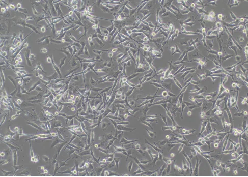
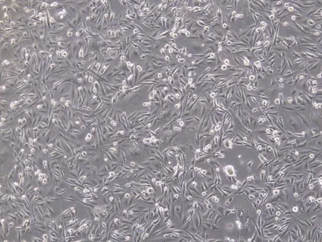
Figure 1: Normal MDA-MB-231 cells
The cells are epithelial-like, with most of the cells spindle-shaped and a few cells round. During the culture process, black spots on the cell bodies are a normal phenomenon.
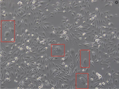
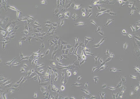
Figure 2: Poor growth status of MDA-MB-231 cells
The cell morphology has changed, and cellular senescence has occurred (as shown in the red box). For example, cells have lost their original spindle-shaped or round morphology and have become irregular or wrinkled.
MDA-MB-231 Cell Culture Protocol
1. Cell Thawing
1) Preparation: Pipet 7 mL of complete medium into a centrifuge tube;
2) Thawing: Take out the cryopreserved vial from the dry ice, hold the cap with forceps, quickly thaw cells in a 37℃ water bath by gently swirling the vial (Note: keep the cap out of the water). In about 1 minute, it would completely thaw;
3) Centrifuge: Transfer the thawed cells to the prepared centrifuge tube by pipette, close the lid, and centrifuge at 1100 rpm for 4 mins, then carefully remove and discard the supernatant;
4) Resuspension and Seeding: Resuspend the cells with 1 mL of fresh complete medium and then transfer to appropriate culture flasks;
5) Cell Culture: Place the culture plates or flasks in a 37℃ incubator and observe the cell status after 24 hours.
2. Cell Passaging (for T25 flask)
1) When the cells are 80%-90% confluent, it is ready to passage. Inside the ultra-cleanbench, remove and discard the medium from the flask and briefly rinse the cell 1-2 times with 5 mL PBS;
2) Add 1 mL of trypsin solution and allow it completely cover the cells, place the flask into the incubator and incubate for 1-2 mins, until the majority of the cells become round and non-adherent as observed under the microscope, a large number of cells detached from each side when gently shaking and tapping the flask, terminate trypsin digestion immediately;
3) Add complete medium to stop digestion, the volume is 2 times of trypsin (i.e. 2 mL for T25 flask). Then transfer the cells into a 15 mL centrifuge tube;
4) Centrifuge at 1100 rpm for 4 mins, then discard the supernatant. Then resuspend the cells with fresh complete medium;
5) Passage the cells at 1:2-1:3 passage ratio and observe the cell status after 24 hours.
3. Cell Cryopreservation
1) Collect the cells: Same as procedures of cell passaging. Digest the cells and transfer to a centrifuge tube;
2) Centrifuge: Centrifuge at 1100 rpm for 4 mins, then discard the supernatant;
3) Resuspension and Cryopreservation: Resuspend the cells with fresh complete medium. Adjust the cells to the required density (1x10⁶ cells/mL) and then transfer to cryovials, labeled with the cell name, source, cell passage number, and date of cryopreservation ;
4) Place the cryovials in 4°C pre-cooled Freezing Container, then put the container in -80℃ freezers. Stay overnight, transfer the cryovials to liquid nitrogen for long-term storage.
Download the MDA-MB-231 cell line culture guide for free now>>>
MDA-MB-231 Cell Culture FAQ
How to Adjust Poor Cell Condition?
- 1. MDA-MB-231 Cell Culture Medium and Serum: Ensure that you are using the correct basalculture medium and adding an appropriate amount of serum; the serum concentration can be adjusted according to the cell condition.
- 2. Cell Culture Environment: Ensure that the culture temperature, humidity, and gas conditions are normal.
- 3. Avoid using expired or long-stored culture media: freshly prepared complete culture media is recommended to be used within two weeks.
- 4. Cell Passage Operations: When passaging cells, pay attention to the digestion time and trypsin concentration to avoid cell damage caused by either too long or too short digestion times.
How to Adjust Cell Senescence?
- 1. Ensure that the culture conditions and environment are correct.
- 2. Avoid excessive trypsin digestion: Use an appropriate trypsin concentration and digestion time, and avoid vigorous pipetting to prevent cell damage.
- 3. Increase serum concentration/select high-quality fetal bovine serum.
- 4. Adjust cell density: High cell density can lead to nutrient deprivation and accumulation of metabolic waste, which accelerates cell senescence. The cell density should be maintained within an appropriate range.
- 5. Add growth factors that can promote cell proliferation and differentiation, which can help improve the state of cell senescence.
- 6. Switch to use the cells with lower passage number.
- 7. Select and purify clones from the senescent cell population using methods like single-cell cloning to isolate cells with growth advantages.
Have more questions? Feel free to contact our experts now>>>
MDA-MB-231 Cell Gene Editing Tips
Precautions for Transfection of MDA-MB-231 Cells:
- 1. Ensure the cell condition is good, and the cells are in the logarithmic growth phase, with a cell density generally around 80-90%.
- 2. Pay attention to the digestion time of the cells to avoid over-digestion, which can cause damageto the cells.
- 3. During the experiment, ensure the cells are thesingle cells by pipetting to avoid cell clumping.
- 4. Cell viability during the experiment should be ≥80%.
Notes for Lentivirus Method:
- 1. During the experiment, the cell confluence should be between 30-40%before infection, and not to be too high.
- 2. Conduct a preliminary experiment before the formal experiment to find the most suitable MOI; add the transfection enhancerPolybrene before infection.
- 3. Ensure that the transfection reagent is well mixed before use to guarantee its uniformity.
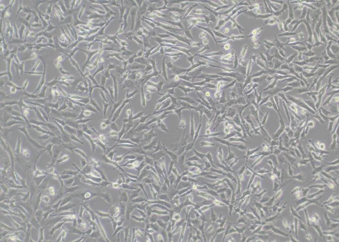
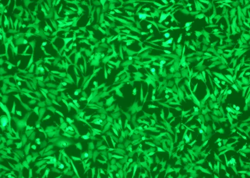
Figure 3: Lentivirus Infection of MDA-MB-231 Cells
Notes for Electroporation Method:
- 1. Control the amount of cells during the experiment, and after electroporation, transfer them onto suitable culture plates.
- 2. Ensure that the cell adhesion rate after electroporation is ≥ 70%.
- 3. Control the experimental time during transfection; the entire electroporation process should not be excessively long.
Single-cell Cloning for MDA-MB-231 Cells:
- 1. Ensure that the cell status is normal and that there are few senescent cells prior to single-cell cloning. It is recommended to maintain the confluence of cells at around 70%.
- 2. During cloning, the cell viability should be ≥85%.
- 3. Conduct a preliminary experiment to find the appropriate cloning dilution gradient to avoid too low a proportion of single clones.
- 4. After diluting the cell count, the results should preferably fall between 1*10^6-2*10^6.
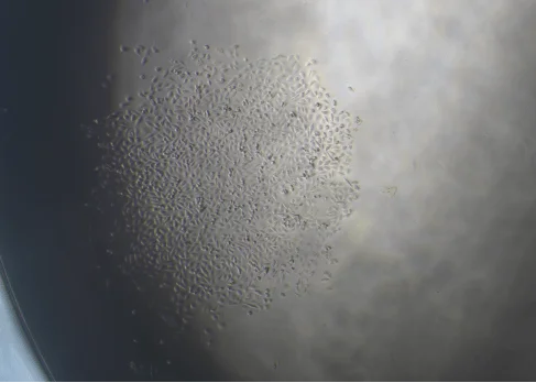
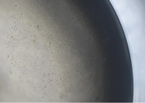
Figure 4: Monoclone of MDA-MB-231 cell
Conclusion
The MDA-MB-231 cell line is widely used in breast cancer metastasis research due to its high invasiveness and epithelial-like morphology. Successfully maintaining cell viability and achieving efficient gene editing requires careful control of digestion time, confluency, and passage frequency. This article offers a complete MDA-MB-231 cell culture protocol, covering thawing, passaging, cryopreservation, lentiviral transduction, electroporation, and single-cell cloning—essential for consistent, reproducible results.
Ubigene offers high-quality, STR-authenticated MDA-MB-231 cells, verified in the ExPASy Cellosaurus database and optimized for both routine culture and gene editing applications. We also provide EZ-editor™ editing services to help you construct custom knockout, knockin, or reporter-tagged MDA-MB-231 models.
Need more authenticated cells for your study? Explore our complete cell bank>>>
 Promotions


