
Commonly Used Cell Lines in Ovarian Cancer Research



Ovarian cancer is one of the deadliest malignancies of the female reproductive system worldwide, characterized by its complex biology, which makes it challenging for researches. To gain deeper insights into its mechanisms, drug responses and resistance, researchers often rely on various in vitro ovarian cancer cell lines. These models provide a solid foundation for researches. This article introduces some commonly used ovarian cancer cell lines and list their applications in gene editing research.
Human Ovarian Cancer Cell Line (OVCAR-3)
Source: Human ovarian adenocarcinoma cell
Characteristics:
This cell line is
sensitive to hormones
, expressing androgen, estrogen, and progesterone receptors. It is
resistant to doxorubicin, melphalan, and cisplatin, making this cell
line a suitable model for studying
drug resistance in ovarian cancer
, aids in evaluating hormone therapies. Besides, this cell line is
highly tumorigenic and is capable of forming tumors in vivo, it is
widely used for studying tumor growth, metastasis, and immune evasion
mechanisms in ovarian cancer research.
Application:
To investigate the role of DCAF13 in ovarian cancer, Tang et al. studied
DCAF13 knockout in OVCAR-3 cells
. They observed significant reductions in proliferation ability of this
knockout cell line. The cell cycle is arrested in G1 phase, DNA damage
mechanism is activated, and migration ability is significantly
inhibited. After transplanting DCAF13 knockout cells into nude mice, the
tumor size and weight were lower than the wild type. And cell cycle,
proliferation, and apoptosis related indicators in the tumor tissue
changed, indicating that tumor in vivo proliferation was inhibited. This
highlights DCAF13's impact on tumor proliferation and growth, providing
valuable insights into ovarian cancer mechanisms and potential
therapeutic targets
[1]
.
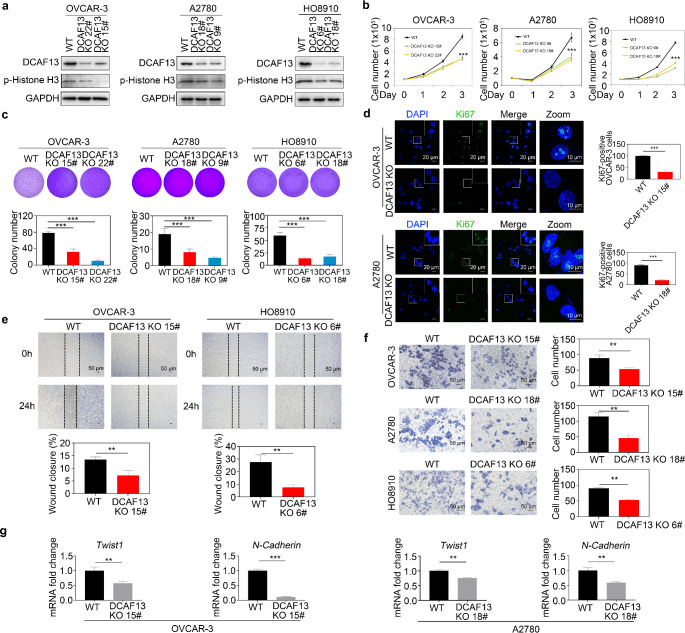
Figure 1: Loss of DCAF13 inhibits proliferation, colony formation, and migration of ovarian cancer cells.
Human Ovarian Adenocarcinoma Cell Line (SK-OV-3)
Source: Human serous cystadenocarcinoma tissue
Characteristics:
This cell line has
relatively high proliferation ability and invasiveness
in vitro culture, and is tolerant to tumor necrosis factor and various
cytotoxic drugs (including diphtheria toxin, cisplatin, and
doxorubicin). Often used in studies on proliferation, invasion,
metastasis, and drug sensitivity of ovarian cancer.
Application:
Using SK-OV-3 cells, Li et al. conducted a
genome-wide CRISPR/Cas9 screening
and identified PCMT1 as a key gene of metastasis and anoikis resistance.
They then take a step further and construct a
PCMT1 knockout in SK-OV-3 cell line
, and confirmed its effects on biological behaviors such as cell
proliferation, migration, adhesion, and invasion, as well as its
pro-apoptotic effect. Moreover, in vivo experiments showed a decrease in
tumor formation, ascites, and metastasis ability after knocking out
PCMT1 gene. The correlation between in vitro and in vivo studies has
strongly demonstrated the importance of PCMT1 in the occurrence,
development, and metastasis of ovarian cancer
[2]
.
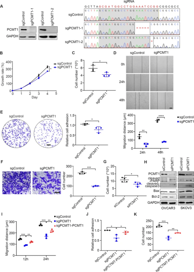
Figure 2: PCMT1 enhances migration, adhesion, and spheroid formation in vitro.
Human Ovarian Adenocarcinoma Cell Line (Caov-3)
Source: Human ovarian papillary serous cystadenocarcinoma
Characteristics:
The genome of Caov-3 cells showed nonsense mutations in the p53 gene and
multiple copies of the ovarian cancer gene PIK3CA. This cell line is
sensitive
to vinblastine, cisplatin, and doxorubicin. Commonly used to study the
mechanisms, gene expression, and signaling pathways of ovarian cancer .
Application:
Chen et al. investigated BRDT's role in ovarian cancer using
BRDT knockout in Caov-3 cells
. The knockout suppressed growth, viability, proliferation, and
migration while inducing apoptosis. Conversely, BRDT overexpression
promoted proliferation, and migration, suggesting that targeting BRDT
could be a potential therapeutic strategy for ovarian cancer
[3]
.
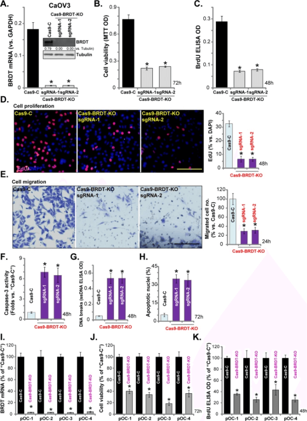
Figure 3: BRDT knockout effectively suppresses ovarian cancer progression.
Human Granulosa Cell Line (KGN)
Source: Human granulosa cell tumor tissue
Characteristics:
This cell line grows slowly and has the ability to synthesize steroid
hormones , expressing functional follicle stimulating hormone receptors.
It is often used to study the pathogenesis, hormone regulation
mechanisms, and discovery of new treatment methods of ovarian granulosa
cell tumors. Meanwhile, it can also be used to study the molecular
mechanisms of ovarian cancer , such as hormone receptor expression,
signaling pathways, etc.
Application:
In order to study the importance of FUT8 in regulating the function of
follicle stimulating hormone receptor (FSHR) in female fertility, Wang
et al. established
FUT8 knockdown (KGN-KD) and FUT8 restored (KGN-KD-Re) cells using
KGN cell line
. By detecting core fucosylation level, FUT8 enzyme activity, FUT8 mRNA
expression, cell proliferation, FSHR expression and related signal
pathways, they found that FUT8 plays an important role in core
fucosylation, cell proliferation, and signal pathways regulated by
FSHR[4].
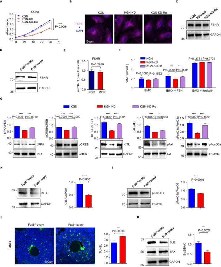
Figure 4: FUT8 knockdown weakens the FSH/FSHR signaling pathway
Summary
These ovarian cancer cell lines, each with unique characteristics, play vital roles in ovarian cancer research. The application of gene editing technology has accelerated the progress of ovarian cancer research. In addition, some studies also use multiple ovarian cancer cell lines for cross validation to enhance the reliability of research results. Researchers can select the most suitable cell lines based on their objectives and experimental conditions.
References
[1]Tang, Ze-Yi et al. “DCAF13 promotes ovarian cancer progression by activating FRAS1-mediated FAK signaling pathway.” Cellular and molecular life sciences : CMLS vol. 81,1 421. 5 Oct. 2024, doi:10.1007/s00018-024-05446-2
[2]Zhang, Jingjing et al. “Genome-wide CRISPR/Cas9 library screen identifies PCMT1 as a critical driver of ovarian cancer metastasis.” Journal of experimental & clinical cancer research : CR vol. 41,1 24. 15 Jan. 2022, doi:10.1186/s13046-022-02242-3
[3]Chen, Ling, et al. "BRDT promotes ovarian cancer cell growth." Cell Death & Disease 11.11 (2020): 1021.
[4]Wang, Tiantong et al. “Core fucosylation regulates the ovarian response via FSH receptor during follicular development.” Journal of advanced research, S2090-1232(24)00038-9. 26 Jan. 2024, doi:10.1016/j.jare.2024.01.025
 Promotions