
CDK10 Loss Sensitizes Cancer Cells to CDK12/13 Inhibitors

Introduction
CDK12 and CDK13 are transcription-associated kinases that regulate transcriptional elongation of DNA damage repair genes, making them promising therapeutic targets in epithelial cancers. However, the lack of reliable predictive biomarkers has limited their clinical application. CDK10 remains relatively understudied. Previous research has linked it to ETS2 degradation, ciliogenesis, and endocrine therapy resistance, but its role in regulating responses to transcription-targeted drugs remains unclear. Through genome-wide CRISPR screening across multiple tumor cell lines , this study identified CDK10 loss as a broadly acting sensitizing factor for CDK12/13 inhibitors. The researchers further uncovered a previously unrecognized role for CDK10 in maintaining transcriptional recovery and establishing a transcriptional buffering barrier, suggesting that CDK10 may serve as a predictive biomarker for patient stratification and a potential target for combination therapy .
Key Research Innovations
- Conceptual Innovation: Multi-cell-line screening demonstrated that CDK10 loss broadly sensitizes tumor cells to CDK12/13 inhibitors . CDK10 maintains RNA polymerase II functional recovery following drug treatment through its kinase activity. Its loss leads to persistent inhibition of long-gene transcription and induces apoptosis. This mechanism is independent of the ETS2 pathway and selectively affects responses to transcriptional kinase inhibitors without altering cisplatin efficacy, supporting the potential use of CDK10 as a predictive biomarker.
- Technical Innovation: Parallel CRISPR screening across multiple cell lines improves the generalizability of target identification. Combined knockout, wild-type rescue, and kinase-dead mutant rescue experiments establish the requirement for CDK10 kinase activity. Time-course RNA-seq and ChIP analyses characterize dynamic transcriptional changes, with CDX models providing in vivo functional validation.
Research Workflow
- 1. Perform parallel CRISPR screening across six tumor cell lines to identify drug-sensitizing genes, including CDK10.
- 2. Conduct knockout and rescue experiments in multiple breast and ovarian cancer cell lines to determine whether CDK10 kinase activity governs drug sensitivity.
- 3. Compare different inhibitors and cisplatin to establish the pathway specificity of the sensitizing effect.
- 4. Apply multi-omics approaches to investigate the molecular mechanisms by which CDK10 loss impairs transcriptional recovery.
- 5. Validate the enhanced antitumor effects in breast and ovarian cancer CDX mouse models.
Key Findings
1. Genome-Wide CRISPR Screening Identifies CDK10 as a Sensitizing Gene for CTX-439
Parallel CRISPR knockout screening was performed across six breast and ovarian cancer cell lines, followed by comparison of sgRNA abundance between drug-treated and control groups. CDK10, FAM58A, and other sensitizing genes were consistently enriched across multiple models. Functional knockout validation demonstrated that loss of FAM58A and TTC14 reduced the drug IC50. Long-term treatment following HNRNPA2B1 knockout significantly suppressed cell proliferation, whereas SCAF4 knockout induced drug resistance, confirming the reliability of the screening system.
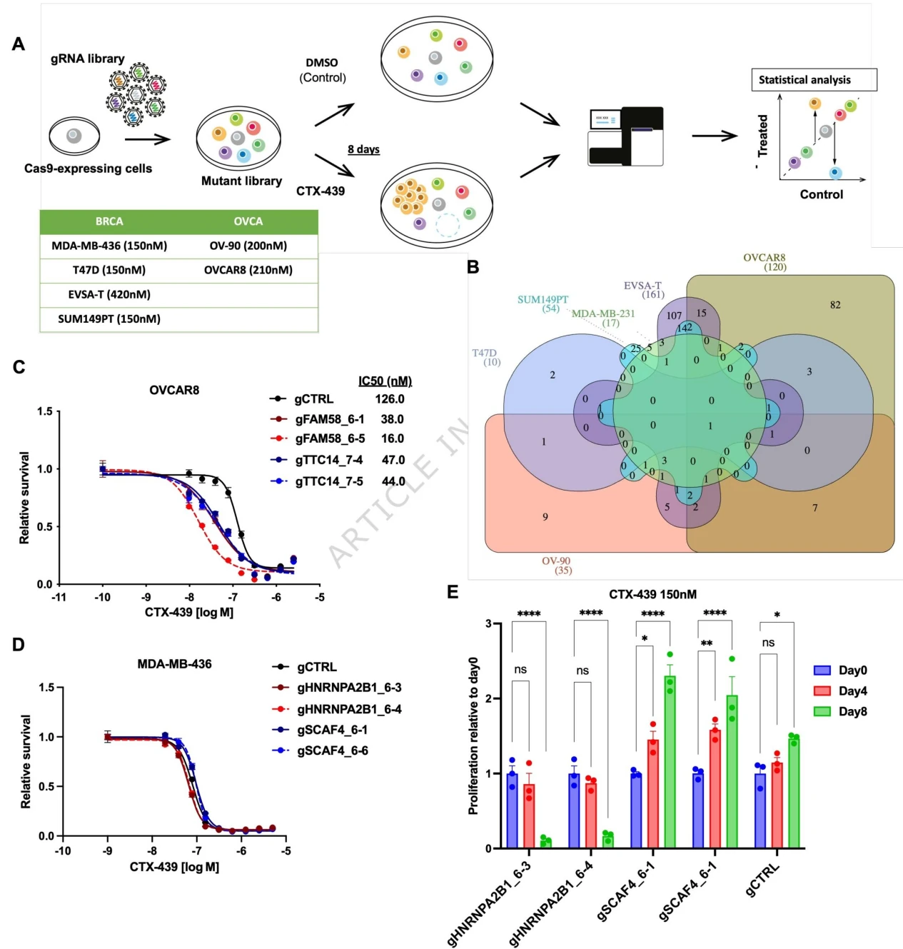
Figure 1. Genome-wide CRISPR-Cas9 screening identifies genetic determinants of CTX-439 sensitivity across multiple cancer models.
2. CDK10 Loss Sensitizes Ovarian Cancer Cells to CDK12/13 Inhibitors
CDK10 was knocked out in three ovarian cancer cell lines: OV-90, OVCAR8, and ES2. CDK10 knockout did not affect baseline cell proliferation but significantly reduced the IC50 of CTX-439 and markedly increased apoptosis. Reintroduction of wild-type CDK10 restored the drug-response phenotype, whereas the kinase-dead mutant failed to do so, demonstrating that the function of CDK10 depends on its kinase activity.
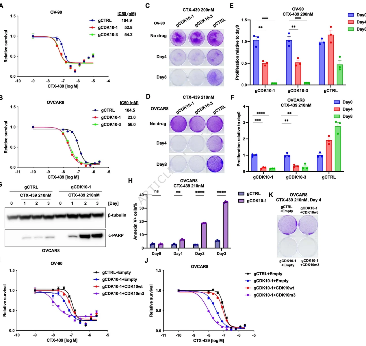
Figure 2. CDK10 knockout enhances the sensitivity of ovarian cancer cells to CTX-439.
3. Breast Cancer Models Confirm the Sensitizing Effect of CDK10 Loss
In MDA-MB-231 and EVSA-T breast cancer cells, CDK10 knockout enhanced drug-induced cell killing and increased apoptosis. The kinase-dead mutant failed to reverse the sensitized phenotype, demonstrating that this regulatory mechanism is also conserved in breast cancer models.
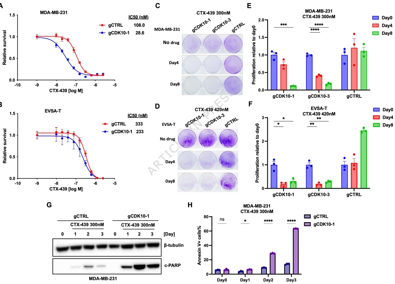
Figure 3. CDK10 loss enhances CTX-439 efficacy in breast cancer models.
4. CDK10 Loss Specifically Sensitizes Cells to CDK12/13 Inhibitors
CDK10-knockout cells exhibited increased sensitivity to both THZ531 and SR-4835. However, no significant differences in cell viability were observed following cisplatin treatment. These findings indicate that the vulnerability caused by CDK10 loss is specific to inhibitors targeting transcriptional elongation rather than representing a broad increase in sensitivity to chemotherapy.
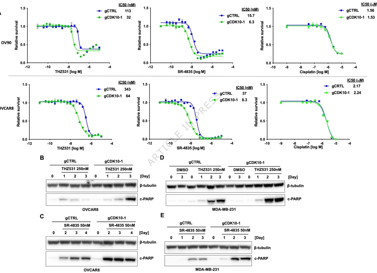
Figure 4. CDK10 selectively regulates cellular responses to CDK12 inhibitors but does not affect responses to the DNA-damaging agent cisplatin.
5. CDK10 Loss Impairs Drug-Induced Transcriptional Recovery
CDK10 loss did not alter baseline ETS2 expression. Following drug treatment, wild-type cells gradually restored RNA polymerase II Ser2 phosphorylation and recovered transcription of long genes such as CAPN7 and SENP6. In contrast, transcriptional suppression persisted in CDK10-knockout cells. Time-course PCA of global transcriptomic profiles further demonstrated that these cells had lost their ability to compensate for drug-induced transcriptional disruption.
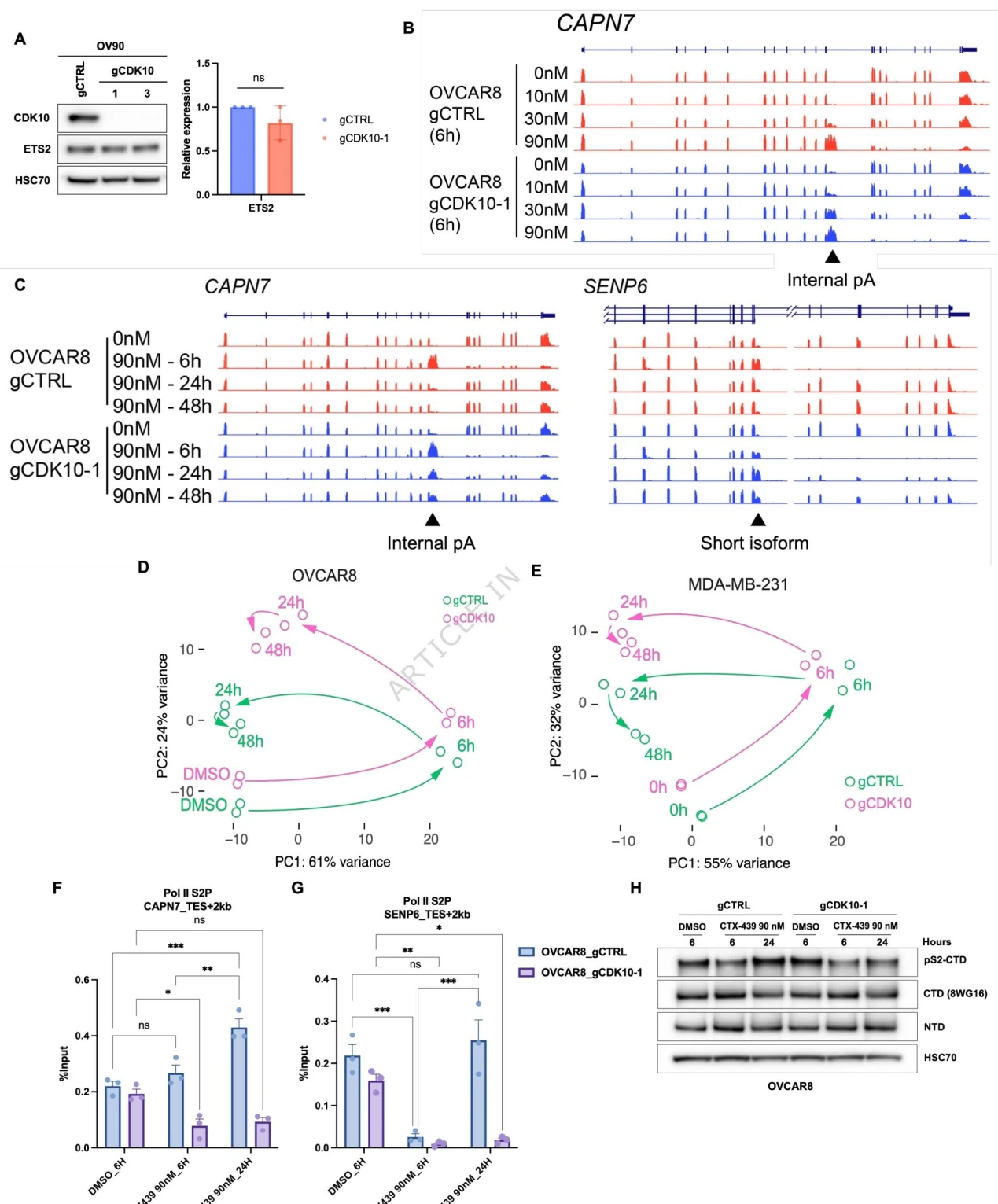
Figure 5. CDK10 supports transcriptional recovery following CDK12 inhibition.
6. In Vivo CDX Models Confirm That CDK10 Loss Enhances the Antitumor Activity of CTX-439
Subcutaneous xenograft models using OVCAR8 and MDA-MB-231 cells were established to evaluate the in vivo effects of CDK10 loss. Following CTX-439 treatment, tumors in the CDK10-knockout groups exhibited significantly greater growth inhibition than those in the control groups. The effect was dose-dependent, providing in vivo confirmation of the sensitizing phenotype observed in vitro.
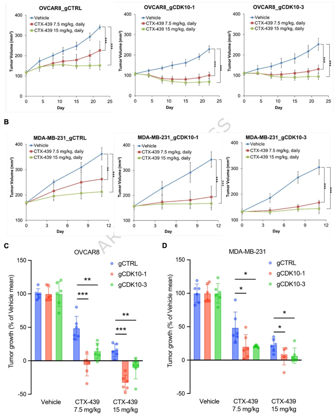
Figure 6. CDK10 knockout enhances the in vivo efficacy of CTX-439.
Conclusion
Through multi-model CRISPR screening , this study identifies CDK10 loss as a shared transcriptional vulnerability in breast and ovarian tumors. As a transcriptional buffering factor, CDK10 relies on its kinase activity to facilitate recovery from transcriptional damage induced by CDK12 inhibition. Its loss results in persistent transcriptional disruption and tumor cell apoptosis. Importantly, this effect is specific to CDK12 inhibitors, and in vivo experiments confirm that CDK10 loss can enhance their therapeutic efficacy. These findings suggest that CDK10 may serve as a predictive biomarker and provide a new rationale for combination strategies targeting transcriptional vulnerabilities in cancer.
Ubigene CRISPR-iScreen™ Libraries
Ubigene has independently developed the CRISPR-iScreen™ technology platform to enable efficient and reliable CRISPR screening. We offer more than 40 ready-to-ship CRISPR plasmid libraries, covering genome-wide knockout, inhibition, and activation libraries for human, mouse, green monkey, and pig species. In addition, our specialized CRISPR knockout, inhibition, and activation sublibraries target genes associated with kinases, the cell cycle, membrane proteins, metabolism, and other biological pathways, helping researchers efficiently identify potential targets. Our portfolio also includes 150+ ready-to-ship CRISPR library virus products and 1,000+ CRISPR library Cell Pool products , as well as comprehensive end-to-end CRISPR library screening services , supporting in vitro and in vivo functional screening, NGS sequencing, and data analysis. Our screening models cover a wide range of cell types, including tumor cells, immortalized cells, immune cells (T cells and NK cells), and human induced pluripotent stem cells (iPSCs), supporting drug target discovery, disease mechanism studies, and functional genomics research.
 Promotions