
VPS34 Drives Cuproptosis Resistance and Radioresistance


Introduction
Cuproptosis, a form of regulated cell death triggered by copper binding to lipoylated proteins in the TCA cycle, such as DLAT, represents a potential therapeutic strategy for overcoming cancer treatment resistance. However, how tumor cells sense copper stress and evade cuproptosis, and how these processes relate to radioresistance, remain poorly understood. Intracellular copper homeostasis depends on the balance between the copper importer SLC31A1 and the copper exporter ATP7A. Through genome-wide CRISPRa screening, this study identified VPS34 as a key negative regulator of cuproptosis . The researchers further demonstrated that PRMT5 methylates VPS34 at R174 and recruits USP10 to promote its deubiquitination and stabilization. Stabilized VPS34 subsequently reduces intracellular copper through two complementary mechanisms: promoting SLC31A1 degradation and facilitating ATP7A translocation to the plasma membrane. This pathway is activated by radiotherapy and contributes to radioresistance, suggesting that targeting it may enhance radiosensitivity.
Key Research Innovations
- Conceptual Innovation: First identification of VPS34 as a key negative regulator of cuproptosis, revealing a dual mechanism of copper transport regulation involving reduced copper uptake and enhanced copper export. The study also elucidates a novel PRMT5-mediated VPS34 R174 methylation-USP10 deubiquitination pathway that stabilizes VPS34, establishing a causal link between disrupted copper homeostasis and radioresistance.
- Technical Innovation: CRISPRa activation screening combined with DIA proteomics identified key effector molecules. Integration of the IP-MS interactome with CCLE drug-sensitivity data identified PRMT5. Mass spectrometry, a site-specific antibody, and the R174K mutant were used to validate the methylation site. Molecular docking characterized the interaction interface, while in vivo experiments evaluated the combination of inhibitors and radiotherapy.
Research Workflow
- Perform genome-wide CRISPRa screening to identify genes associated with cuproptosis resistance, including VPS34, and investigate the protective role of autophagy.
- Elucidate how VPS34 maintains copper homeostasis through SLC31A1 degradation and ATP7A trafficking.
- Integrate IP-MS and CCLE datasets to identify the upstream methyltransferase PRMT5 and validate symmetrical dimethylation of VPS34 at R174.
- Investigate how PRMT5 recruits USP10 to suppress K48-linked ubiquitination and stabilize VPS34.
- Use R174K mutant rescue experiments to establish the functional requirement for methylation, followed by in vitro and in vivo validation of the PRMT5–VPS34 axis in radioresistance and radiosensitization.
Key Findings
1. Genome-Wide CRISPRa Screening Identifies VPS34 as a Negative Regulator of Cuproptosis
Researchers first performed genome-wide CRISPR activation (CRISPRa) screening in cells treated with the copper ionophore elesclomol-copper (ES-Cu) and validated the reliability of the screening system. The results confirmed the robustness of the screening approach. Copper export factors (ATP7A and MT2A/B) were enriched as positive hits, whereas cuproptosis effectors (FDX1 and DLAT) and SLC31A1 were identified as negative hits. Top-ranked hits were enriched in pathways associated with autophagy and endosomal membrane trafficking. DIA proteomics identified VPS34 as the protein showing the most pronounced increase in expression. Western blotting confirmed that ES-Cu induced VPS34 expression in a dose-dependent manner, while FYVE-probe analysis demonstrated increased PI3P production. VPS34 overexpression conferred resistance to cuproptosis, whereas VPS34 knockout increased cellular sensitivity and exacerbated DLAT oligomerization (an effect that could be reversed by FDX1 knockdown).
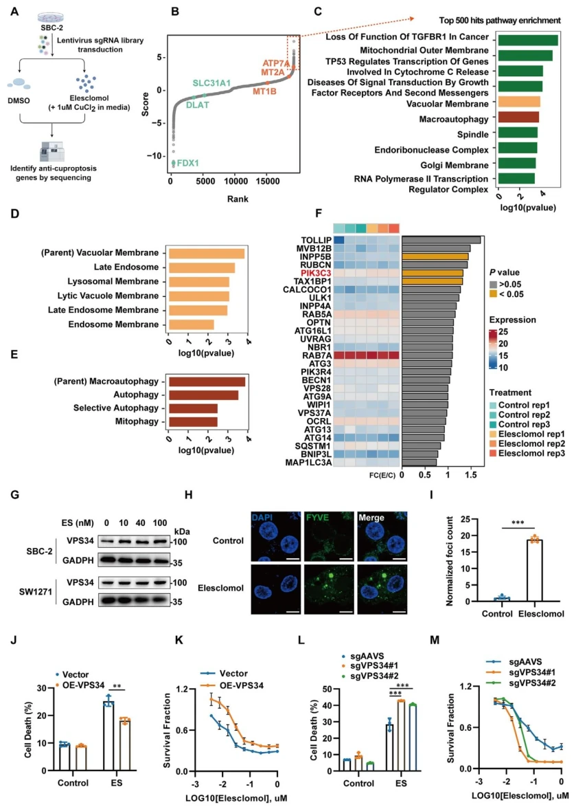
Figure 1. Genome-wide CRISPRa screening identifies VPS34 as a negative regulator of copper-induced cell death.
2. VPS34 Maintains Copper Homeostasis by Coordinating SLC31A1 Degradation and ATP7A Trafficking
VPS34 overexpression significantly reduced intracellular copper levels, as demonstrated by flow cytometry using the CS1 probe and confocal microscopy, whereas VPS34 knockout resulted in substantial copper accumulation. Mechanistically, VPS34 overexpression did not alter total ATP7A protein levels but significantly reduced SLC31A1 protein abundance. The lysosomal inhibitor bafilomycin A1 (BafA1) blocked this degradation, confirming that VPS34 accelerates SLC31A1 turnover through the lysosomal pathway to reduce copper uptake. Regarding ATP7A, VPS34 overexpression significantly promoted its translocation to the plasma membrane. Colocalization analysis revealed increased colocalization of ATP7A with RAB7A (a late endosomal marker), but no significant overlap with EEA1 (an early endosomal marker), LAMP1 (a lysosomal marker), or LC3 (an autophagosomal marker). These findings indicate that VPS34 promotes ATP7A recycling to the plasma membrane through RAB7A-positive endosomal sorting, bypassing degradative compartments and thereby enhancing copper export.
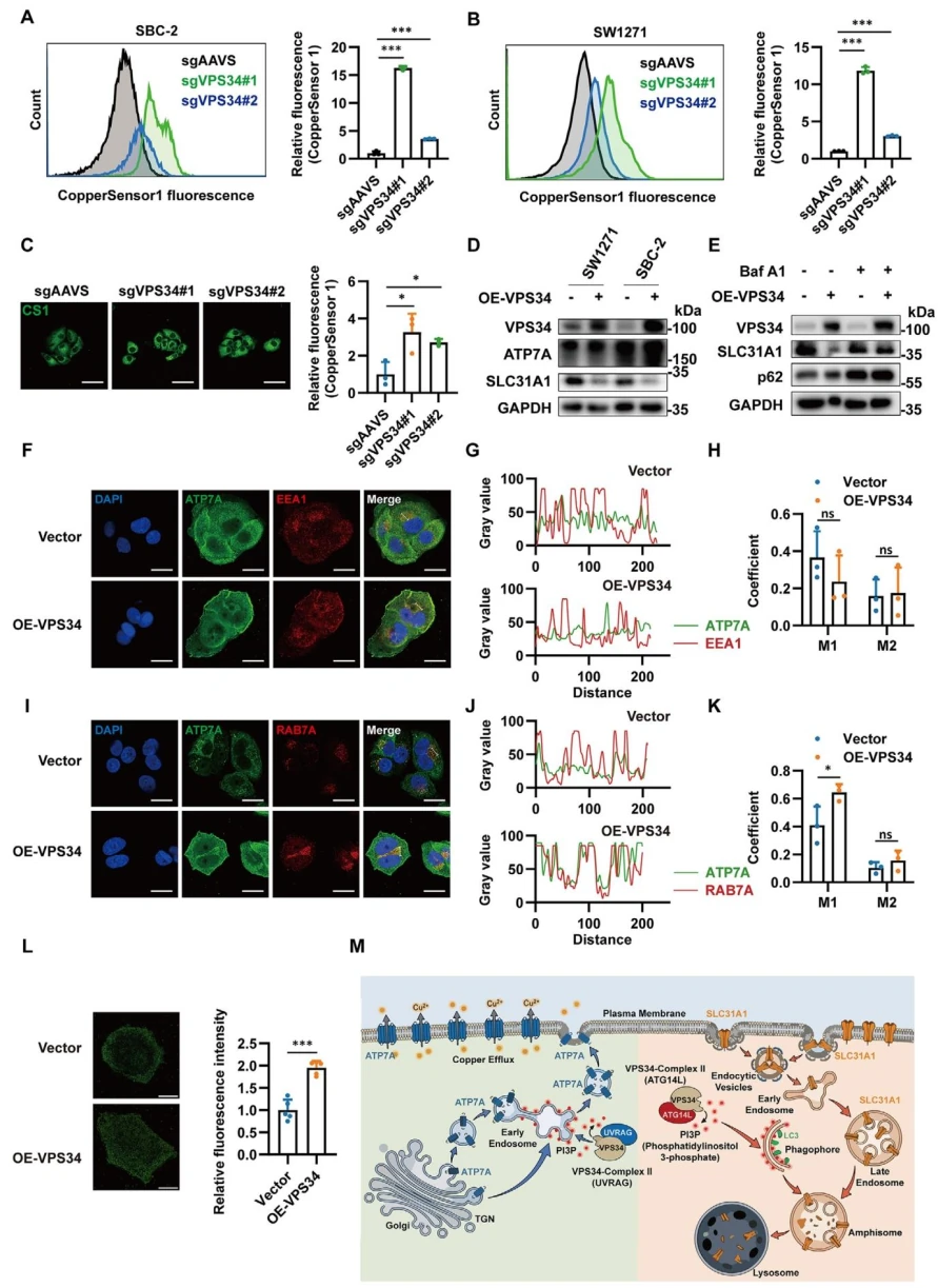
Figure 2. VPS34 maintains intracellular copper homeostasis by coordinating SLC31A1 degradation and ATP7A trafficking.
3. PRMT5 Interacts with VPS34 and Mediates Symmetrical Dimethylation at R174
Researchers subsequently identified PRMT5 by intersecting the VPS34-interacting proteins identified through IP-MS (694 proteins) with proteins associated with elesclomol sensitivity in the CCLE dataset. PRMT5 expression was also upregulated in a dose-dependent manner following ES-Cu treatment. Co-immunoprecipitation (Co-IP) confirmed the interaction between PRMT5 and VPS34 and mapped the interaction to the C2 domain of VPS34. An antibody against symmetrical dimethylarginine (SDMA) demonstrated that VPS34 undergoes symmetrical dimethylation, which was positively regulated by PRMT5. Mass spectrometry identified R174 as the methylation site, a residue conserved across vertebrates. The R174K mutant showed reduced methylation. A site-specific antibody and molecular docking analysis further validated this finding. Molecular docking showed that R174 is positioned within the PRMT5 catalytic pocket, with an approximate distance of 5 Å between the S-adenosylmethionine (SAM) group and the guanidino group.
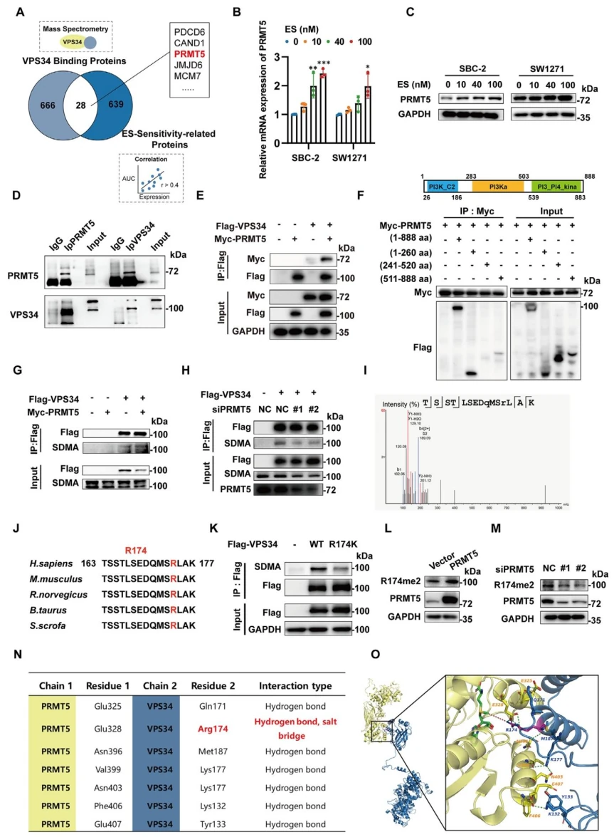
Figure 3. PRMT5 catalyzes SDMA modification of VPS34 at R174.
4. PRMT5 Stabilizes VPS34 by Recruiting USP10 to Suppress K48-Linked Polyubiquitination
Researchers found that PRMT5 upregulated VPS34 protein levels without affecting its mRNA expression, suggesting a post-translational regulatory mechanism. MG132, but not BafA1, prolonged the half-life of VPS34. Cycloheximide (CHX) chase assays further confirmed that PRMT5 overexpression extended VPS34 stability, whereas PRMT5 knockdown or the R174K mutation accelerated its degradation. VPS34 was found to undergo predominantly K48-linked polyubiquitination. PRMT5 overexpression suppressed this modification, whereas the R174K mutant exhibited increased ubiquitination. Among seven candidate deubiquitinating enzymes, siRNA screening identified USP10 as the key mediator. Co-IP confirmed the interaction between USP10 and VPS34, and PRMT5 enhanced this interaction. Together, these findings demonstrate that R174 methylation promotes USP10 recruitment, leading to VPS34 deubiquitination and stabilization .
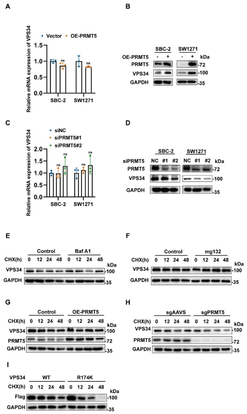
Figure 4. SDMA modification of VPS34 prevents its degradation and promotes autophagy.
5. R174 Methylation Is Essential for VPS34 Stability and Cellular Resistance to Cuproptosis
PRMT5 overexpression reduced intracellular copper levels, promoted lysosomal degradation of SLC31A1, and enhanced ATP7A translocation to the plasma membrane and its colocalization with RAB7A. Functionally, these changes increased resistance to cuproptosis. Conversely, PRMT5 knockout produced the opposite effects and exacerbated DLAT oligomerization, which could be reversed by FDX1 knockdown. In subcutaneous xenograft models, tumors in the shPRMT5 group exhibited significantly reduced growth and tumor weight compared with controls. Genetic rescue experiments further established the functional importance of R174 methylation. Reintroduction of wild-type VPS34 into VPS34-knockout cells restored copper export and resistance to cuproptosis. In contrast, the R174K mutant was unstable, failed to reduce intracellular copper accumulation, and could not rescue the cuproptosis-sensitive phenotype, confirming that R174 methylation is essential for VPS34 function .
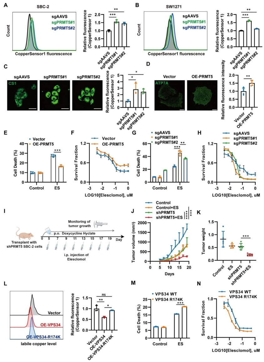
Figure 5. R174 methylation is essential for VPS34 stability and cellular resistance to cuproptosis.
6. The PRMT5–VPS34 Axis Promotes Radioresistance, and Targeting This Pathway Enhances Radiosensitivity
Researchers found that radiotherapy increased intracellular copper levels in a dose-dependent manner, as measured by ICP-MS. ES-Cu enhanced radiosensitivity, whereas copper chelation with TTM had a protective effect. Radiation exposure also increased PRMT5 and VPS34 expression, together with R174 dimethylation. Clonogenic survival assays showed that VPS34 overexpression reduced radiosensitivity, whereas its knockout enhanced radiosensitivity. The R174K mutant failed to rescue the radioresistant phenotype. In vivo experiments further demonstrated that combining radiotherapy with either VPS34 or PRMT5 inhibitors significantly suppressed tumor growth. Clinical analyses revealed that high PRMT5 and VPS34 expression was associated with poor survival outcomes in lung cancer. TCGA data further indicated that patients with high PRMT5 expression exhibited poorer responses to radiotherapy.
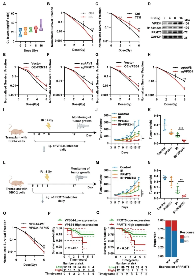
Figure 6. The PRMT5–VPS34 axis contributes to radioresistance.
Conclusion
This study identifies the PRMT5–VPS34 axis as a central pathway underlying cuproptosis escape and radioresistance in cancer . Radiation exposure and copper stress upregulate PRMT5, which methylates VPS34 at R174 and recruits USP10 to promote deubiquitination and stabilize VPS34. Through PI3P signaling, VPS34 simultaneously drives SLC31A1 lysosomal degradation and ATP7A translocation to the plasma membrane, reducing intracellular copper levels through decreased uptake and enhanced export. This mechanism suppresses cuproptosis and compromises radiotherapy efficacy. Targeting PRMT5 or VPS34 may restore copper-induced cytotoxicity and enhance radiosensitivity. PRMT5 inhibitors have already entered clinical development, offering promising translational potential. However, the study has several limitations. The mechanistic findings are primarily based on small-cell lung cancer models, particularly SBC-2 cells. Whether the mechanism is broadly applicable to tissues such as the liver, where ATP7B plays a dominant role in copper export, remains to be established. In addition, the upstream mechanisms through which radiotherapy induces PRMT5, potentially involving ROS-mediated transcription factor signaling, have not been fully elucidated.
Ubigene EZ-iScreen™ Libraries
Ubigene has independently developed the EZ-iScreen™ technology platform to enable efficient CRISPR screening. We offer more than 40 ready-to-ship CRISPR plasmid libraries, covering genome-wide knockout, inhibition, and activation libraries for human, mouse, green monkey, and pig species. In addition, our specialized CRISPR knockout, inhibition, and activation sublibraries target genes associated with kinases, the cell cycle, membrane proteins, metabolism, and other biological pathways, helping researchers efficiently identify potential targets. Our portfolio also includes 150+ ready-to-ship CRISPR library virus products, 1,000+ CRISPR library Cell Pool products, and comprehensive one-stop CRISPR library screening services , supporting in vitro and in vivo functional screening, NGS sequencing, and data analysis. Our screening models cover a wide range of cell types, including tumor cells, immortalized cells, immune cells (T cells and NK cells), and human induced pluripotent stem cells (iPSCs), supporting drug target discovery, disease mechanism studies, and functional genomics research.
 Promotions