
Expert Insights | Practical Tips for AC16 Cell Culture and Gene Editing

Why is your gene editing efficiency in AC16 cells consistently lower than expected? One factor that researchers often overlook is the condition of the cells themselves, which is a critical prerequisite for successful gene editing. In this guide, we take an in-depth look at the key factors affecting AC16 cell culture, along with practical culture strategies and gene editing tips to help you overcome common barriers to editing efficiency.
The AC16 cell line originated from the ventricular tissue of the adult human heart. To establish this immortalized cell line, researchers fused primary ventricular cells with uridine-auxotrophic fibroblasts carrying the SV40 viral gene and lacking mitochondrial DNA. The fibroblasts provided the cells with unlimited proliferative potential. Under in vitro culture conditions containing mitogens, AC16 cells proliferate rapidly. When transferred to mitogen-free medium, they can effectively differentiate into functional, mature cardiomyocytes. As a result, AC16 has become an important cellular model for studying cardiomyocyte lineage differentiation, localized cardiac inflammatory responses, ischemic or chemical injury, and the cardiac safety of candidate drugs.
1. Basic Information on Human Cardiomyocytes (AC16)
- Cell Name: Human Cardiomyocytes (AC16)
- Cell Morphology: Fibroblast-like, adherent
- Culture Medium: 90% DMEM/F12 + 10% FBS
- Atmosphere: 95% air, 5% CO₂
- Temperature: 37°C
- Medium Change: Every 2–3 days
- Split Ratio: 1:3–1:4
Reference Cell Growth Conditions
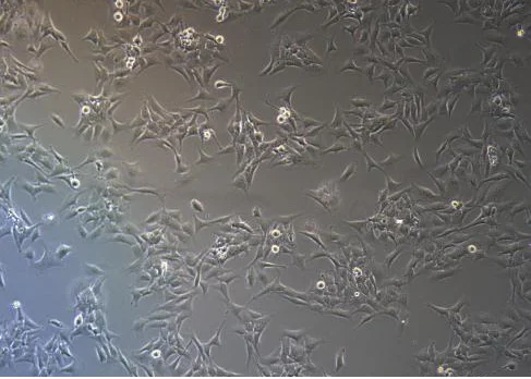
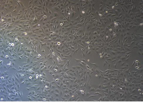
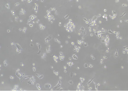
Normal condition: Cells grow adherently. Their morphology is heterogeneous, with elongated spindle-shaped or irregular polygonal cells. Cell boundaries are clear, and cells spread evenly across the culture surface. (See image below.)
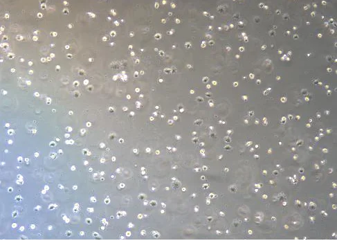
Abnormal condition: Cells lose their characteristic morphology and become shrunken and rounded. Their edges become rough and indistinct, and their refractive properties decrease, making the cells appear darker under the microscope. Cells may also detach and die, or become abnormally enlarged. (See image below.)
2. AC16 Cell Culture
Key Culture Considerations
- Cell condition: The optimal time for passaging is when cells reach 80–90% confluence. For actively growing cells, replace the complete culture medium every 2–3 days.
- Strictly follow the basic culture conditions: Ensure that the cell culture environment is properly maintained and that the correct culture system is used.
- Medium storage: Store the culture medium at 4°C and protected from light, and use it before the expiration date.
- Handling details: Pre-warm the culture medium and trypsin to 37°C before use to minimize temperature stress.
- Use high-quality fetal bovine serum: High-quality FBS is recommended for AC16 cell culture.
- Pay attention to digestion time: Stop digestion immediately when cells become rounded and the spaces between cells begin to increase under the microscope. This helps prevent over-digestion.
Common AC16 Cell Culture Problems and Solutions
1. What Should You Do If the Cells Grow Slowly?
Symptoms: After passaging, the cells require more than 4–5 days to reach 80–90% confluence. Under normal conditions, AC16 cells should reach confluence approximately 2–3 days after passaging.
-
Possible causes:
- Seeding density is too low.
- Poor-quality serum.
- Culture medium issues, such as inappropriate pH or nutrient composition.
-
Solutions:
- Maintain a split ratio of 1:3–1:4 and avoid seeding the cells at an excessively low density.
- AC16 cells are sensitive to serum quality, so use high-quality FBS for culture.
- Confirm that the correct DMEM/F12 (1:1) basal medium is being used and replace the medium regularly every 2–3 days.
2. What Should You Do If the Cell Morphology Is Abnormal?
Symptoms: Cells become shrunken and rounded, develop intracellular vacuoles, or become abnormally enlarged.
-
Possible causes:
- Excessive passaging.
- Improper cell digestion.
-
Solutions:
- Use low-passage cells for experiments.
- Handle cells gently during digestion and pipetting. Avoid over-digestion, terminate digestion promptly, and add a sufficient volume of serum-containing culture medium.
3. What Should You Do If Cell Recovery Is Poor?
Symptoms: Very low cell viability and extensive cell death after thawing.
-
Possible causes:
- Improper thawing: thawing too slowly can cause ice crystal damage.
- Low seeding density: too few cells may result in insufficient autocrine growth factors.
- Poor initial cell condition: cells may have been at an excessively high passage number or in poor condition before cryopreservation.
-
Solutions:
- Follow the rapid-thawing principle: rapidly thaw the cryovial in a 37°C water bath while gently shaking. Complete thawing within 1–2 minutes and remove the vial when only a small amount of ice remains.
- After recovery, seed the cells at approximately 60–80% density whenever possible to support cell growth.
- Consider thawing another vial of cells with a lower passage number.
3. AC16 Gene Editing
Transfection Tips
1. Cell Condition Requirements
- Ensure that the cells are in good condition and use cells in the logarithmic growth phase, with approximately 70–80% confluence.
- Cell viability should be >85%, which can be assessed using trypan blue staining.
- Use low-passage cells.
- Pay close attention to digestion time and avoid over-digestion, which can damage the cells.
- During the experiment, pipette the cells into a single-cell suspension and avoid cell clumping.
2. Transfection Reagents and Preliminary Optimization
- Mix the transfection reagent thoroughly before use to ensure uniformity.
- A preliminary drug-selection experiment is recommended to determine the optimal selection concentration before performing post-transfection selection.
3. Electroporation
- Control the number of cells and seed the electroporated cells into an appropriate culture plate based on the cell number.
- Use a gentle trypsinization procedure and completely neutralize digestion with serum-containing culture medium.
- Wash the cells with PBS 1–2 times to thoroughly remove residual serum and minimize ionic interference during electroporation.
- Perform preliminary experiments to optimize the electroporation parameters.
- Ensure a post-electroporation cell attachment rate of ≥60%.
- Control the experimental duration; the entire electroporation procedure should not be unnecessarily prolonged.
4. Lentiviral Transduction
- Perform preliminary experiments to determine the optimal MOI (multiplicity of infection).
- Maintain cell confluence at 30–40% before viral transduction and avoid excessive confluence.
- Add the transduction enhancer Polybrene before infection.
- Replace the culture medium 24 hours after infection.
- Avoid repeated freeze-thaw cycles of the viral preparation.
- If the transduction efficiency is too low, a second round of infection may be performed, provided that the cells have good tolerance to the virus. Centrifugation-assisted infection can also be considered.
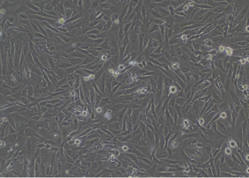
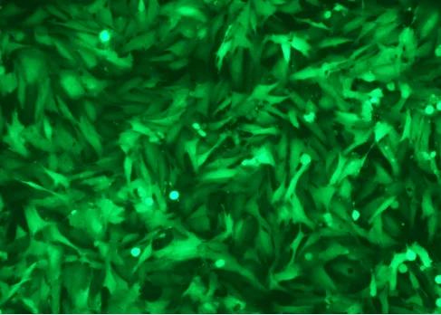
AC16 cells during gene editing using the lentiviral method (left: bright-field; right: fluorescence)
Tips for Single-Clone Experiments
1. Cell Condition Requirements
- Use cells in the logarithmic growth phase for clonal plating. Cell confluence is recommended to be approximately 70% before plating.
- Cell viability should be ≥80% at the time of clonal plating.
2. Reagents and Preliminary Optimization
- Pre-warm all reagents, including culture medium and PBS.
- Gentle dissociation reagents, such as TrypLE Express, are recommended.
3. Plating Strategy
- Perform preliminary experiments to determine an appropriate clonal plating dilution range and avoid an excessively low proportion of single-cell clones.
- When seeding cells into a 96-well plate, ensure that the cells are distributed evenly. Add PBS to the outer wells of the 96-well plate to minimize evaporation.
4. Dilution Method
- Use the limiting dilution method for clonal plating.
- After dilution and cell counting, the cell count is preferably maintained between 1 × 10⁶ and 2 × 10⁶.
If you are planning to use AC16 cells for your research, visit the Ubigene Red Cotton OmniCell Bank to search for Human Cardiomyocytes (AC16). Ubigene offers more than 1,000 wild-type cell lines covering a wide range of research areas. We have also successfully generated gene knockout cells, Luc stable cell lines, Cas9 stable cell lines, and other engineered AC16 cell models to support a wide range of research needs.
 Promotions