New Tumor Suppressor Gene ECHS1 Identified: Bridging the Gap in Early Malignant Transition of Hepatocellular Carcinoma


Introduction
Hepatocellular carcinoma (HCC) is a leading cause of cancer-related death worldwide. Most cases arise from dysplastic nodules (DNs, premalignant lesions) within the context of cirrhosis, with approximately 30% of DNs progressing to malignancy. Recently, the research team led by Lijian Hui at the Center for Excellence in Molecular Cell Science, Chinese Academy of Sciences, published a landmark study in Cancer Cell . Their study focuses on the malignant transformation from precancerous DNs to very early HCC (veHCC). Utilizing evolutionarily related nodule-in-nodule samples , multi-omics sequencing, and spatial transcriptomics, the study reveals core mechanisms: TERT alterations initiate carcinogenesis, copy number alterations (CNAs) drive malignant transformation, and a dual evolutionary mode exists involving immune desert and immune escape phenotypes. This work revises the traditional view that HCC originates from chronic inflammation and provides novel targets for early diagnosis and treatment of liver cancer.
Background
HCC predominantly develops from dysplastic nodules (DNs) in a cirrhotic liver, yet the critical genetic and immune mechanisms driving this early malignant transition remain elusive. Previous studies have largely relied on evolutionarily unrelated precancerous and cancerous tissues, which fails to accurately delineate the authentic trajectory of malignant transformation. Furthermore, while it is widely accepted that HCC originates within a chronic inflammatory microenvironment, the specific immune characteristics of precancerous lesions and the key genomic events driving malignant transformation remain poorly defined.
Objectives
This study aims to characterize the genomic and microenvironmental landscape governing the malignant transition from DN to veHCC using evolutionarily related samples, identifying the molecular drivers and potential targets for early diagnosis and intervention.
Methods
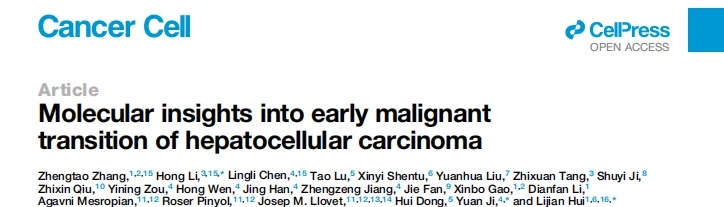
- Clinical Sample Screening: Screened 44,714 FFPE liver specimens over 9 years to acquire 17 evolutionarily related nodule-in-nodule samples (DN + paired veHCC) from 16 patients. An additional 19 cancer-undetermined DNs were included as controls.
- Multi-omics Sequencing: Whole-genome sequencing (WGS, ~35×), RNA-seq, spatial transcriptomics (Visium HD), and multiplex immunofluorescence/immunohistochemistry (IHC).
- Genetic Manipulation and Functional Validation: Human liver organoid models, CRISPR knockout/overexpression, and EdU proliferation assays.
- Bioinformatics Analysis: Mutational signatures, copy number alterations (CNA), cell type deconvolution (MCP-counter), cell-cell communication analysis (SOAPy), and phylogenetic tree construction (Pairtree).
Research Workflow
- Sample Identification: Confirmed that the nodule-in-nodules are evolutionarily related DNs and veHCCs.
- Precancerous Driver Screening: Compared cancer-prone and cancer-undetermined DNs, identifying TERT alteration as the pivotal initiating event.
- Malignant Transformation Drivers: Established that the accumulation of CNAs, rather than SNVs, serves as the primary engine for the DN-to-veHCC transition.
- Immune Landscape Profiling: Revealed that DNs exhibit an "immune-desert" phenotype, whereas a subset of veHCCs presents an "inflamed" + immune evasion phenotype.
- Spatial Mapping: Visualized the spatial distribution of immune and stromal cells utilizing spatial transcriptomics.
- Model Proposal: Summarized a dual evolutionary trajectory characterized by a CNA-driven mode and an immune-evasion mode.
Key Results
1. Identification of Evolutionarily Related DN and veHCC Samples
The study found that 17 cancer-prone DNs and their 21 paired veHCCs shared 7% to 85% of somatic SNVs, confirming a direct phylogenetic relationship. TERT alterations (promoter mutations, HBV integration, or copy number gains) were present in 82% of cancer-prone DNs, which is significantly higher than in cancer-undetermined DNs (32%). While TERT alterations upregulated TERT expression, progressive telomere shortening was observed, indicating that TERT alterations initiate carcinogenesis rather than reverse telomere attrition.
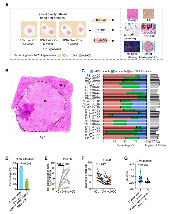
Figure 1. Sample collection and characterization
2. CNA is strongly associated with malignant transition
During the DN-to-veHCC transition, the number of protein-altering SNVs did not significantly increase, however, the CNA burden was markedly elevated. veHCC specifically exhibited loss of chromosome 1p (which harbors tumor suppressor genes such as ARID1A and NR0B2), accompanied by increased chromosomal instability (CIN). Functional validation demonstrated that the knockout of ECHS1 or FGA significantly promoted the proliferation of liver organoids, confirming their roles as early tumor suppressor genes.
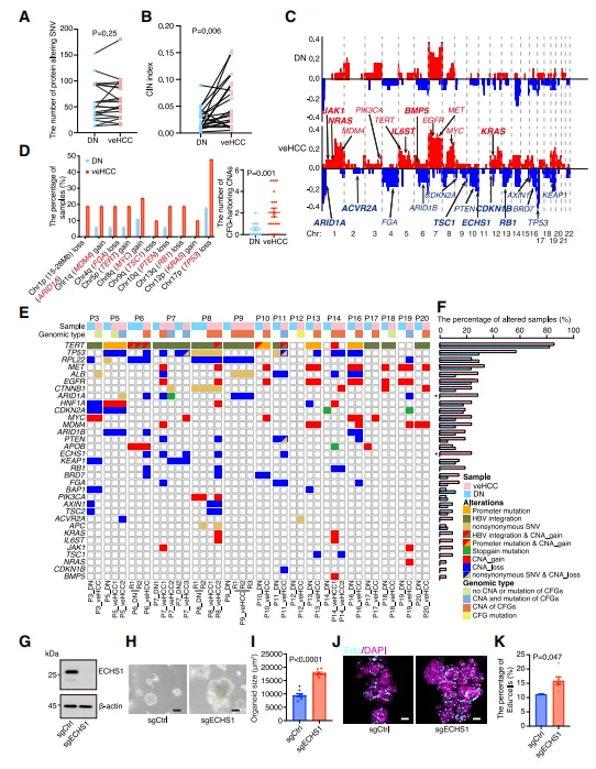
Figure 2. Key genomic alterations driving malignant transition of DNs
3. Precancerous DNs Exhibit an "Immune-Desert" Phenotype
Cancer-prone DNs demonstrated a comprehensive downregulation of inflammatory pathways (e.g., TNFα/NF-κB, IL6/JAK/STAT3), along with decreased expression of type I interferons, chemokines, and TLR receptors. Immune cell infiltration was significantly reduced: macrophages, CD4+/CD8+ T cells, fibroblasts, and endothelial cells were all lower compared to non-cancerous adjacent tissues. Conclusion: HCC originates from an immunologically inert microenvironment, rather than chronic inflammation, fundamentally challenging the traditional paradigm.
Figure 3. Immune-desert phenotype in precancerous DNs
4. "Inflamed" veHCCs Simultaneously Manifest Immune Activation and Evasion
Approximately 43% of veHCCs were classified as the "inflamed" subtype: characterized by high infiltration of T cells and dendritic cells (DCs), but a low CNA burden. Inflamed veHCCs exhibited high expression of PD-1/PD-L1/CTLA-4 , Treg cell enrichment, and prominent T cell exhaustion signatures. Concurrently, oncogenic pathways such as IL6, TNFα, EMT, and KRAS were activated, indicating a synchronous process of immune evasion and malignant progression.
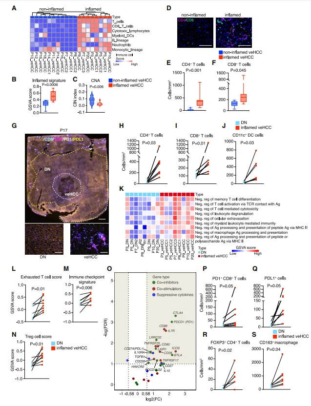
Figure 4. Adaptive immune activation and evasion mechanisms in a subset of veHCCs
5. Spatial Transcriptomics Reveals the Architecture of the Immunosuppressive Microenvironment
Spatial mapping highlighted close interactions between CD8+ T cells and CD4+ T cells, cancer-associated fibroblasts (CAFs), and macrophages. CAFs mediated immunosuppression via the TGF-β signaling pathway, and regions co-localized with CD8+ T cells displayed pronounced immune exhaustion. A subset of veHCCs formed tertiary lymphoid structures (TLS), which correlated with the immune-evasion mode of malignant transformation.
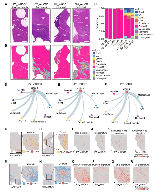
Figure 5. Spatial transcriptomic analysis of immune/stromal cell infiltration, cell-cell communication, and characteristic activation
6. Proposal of a Dual Evolutionary Model for Early HCC Malignant Transition
Based on genomic alterations and immune microenvironment features, the study proposes two parallel evolutionary scenarios for early HCC malignant transition. The first is a CNA-driven progression, characterized by high chromosomal instability, high CNA burden, and low immune infiltration, with TP53 aberrations enriched in this scenario. The second is an inflamed progression coupled with immune evasion, characterized by a low CNA burden, high immune infiltration, and the early establishment of immune evasion programs; in some cases, no additional driver mutations were acquired. Furthermore, both evolutionary modes can coexist within the same patient, demonstrating that tumor heterogeneity is established at the very inception of hepatocarcinogenesis.
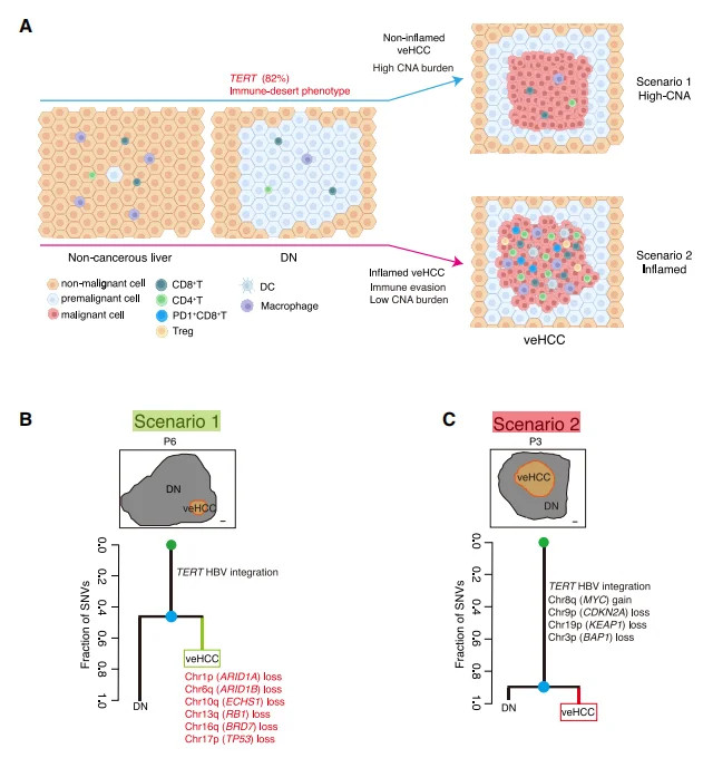
Figure 6. Evolutionary scenarios of malignant transition of DNs
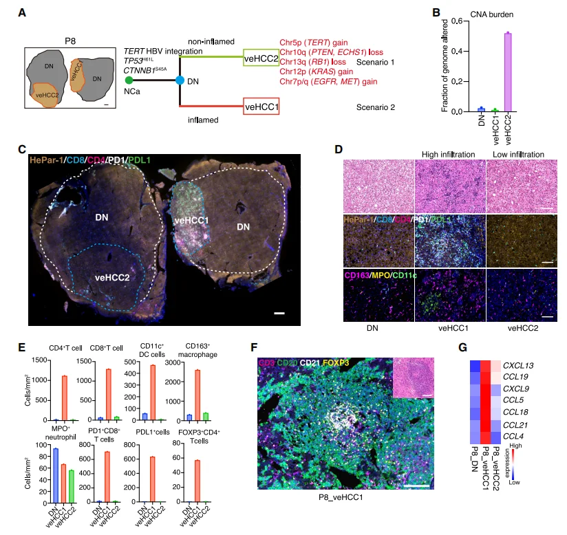
Figure 7. Evolutionary scenario of P8
Significance and Innovations
- Sample Innovation: Pioneered the use of evolutionarily related nodule-in-nodule samples to accurately map the trajectory of early HCC malignant transition.
- Mechanistic Breakthrough: Demonstrated that HCC originates from an immune desert rather than chronic inflammation, identifying TERT alterations as the initiating event and CNAs as the core driver of malignant transformation.
- Subtyping Paradigm: Proposed a dual evolutionary model, providing a strong rationale for early diagnostic stratification and targeted/immunotherapy.
- Target Discovery: Identified ECHS1 and FGA as early tumor suppressor genes, and highlighted TGF-β and immune checkpoints as viable targets for early intervention.
Summary
By leveraging evolutionarily related very early HCC samples, this study reveals the principles of early malignant transformation in HCC—initiated by TERT alterations, driven by CNAs, and shaped by immune microenvironment remodeling. The proposition of a CNA-driven and immune-evasion dual evolutionary model revises traditional theories of HCC origin and provides a novel molecular foundation and strategic direction for early screening, diagnosis, and precision intervention in liver cancer.
Ubigene Biosciences has consistently adhered to the core belief of "Making Gene Editing Easier," continuously iterating our products and services. To date, we has successfully delivered over 13,000 gene-editing cases and possesses over 11,000 cell products (including 8,000+ KO cell lines). Through self-developed innovative technologies, our gene-editing efficiency has been increased by 10 to 20 times compared to traditional methods. To date, Ubigene Biosciences has provided high quality gene-editing services and products to over 10,000 life science laboratories, pharmaceutical companies, and CROs.
The protein encoded by the ECHS1 gene plays a pivotal role in cellular metabolism. Specifically, this protein is a short-chain enoyl-CoA hydratase, primarily involved in the β-oxidation of fatty acids. Fatty acid β-oxidation is a critical pathway for cellular energy production, meaning the function of the ECHS1 gene is directly linked to the cellular energy supply. If you are interested in conducting research on ECHS1 , Ubigene Biosciences can provide ECHS1 knockout cell lines, including lines such as Hep G2, HuH-7, and LoVo. If you have customize gene-editing needs, please feel free to inquire!
Contact us to learn more details >>>> Promotions


