
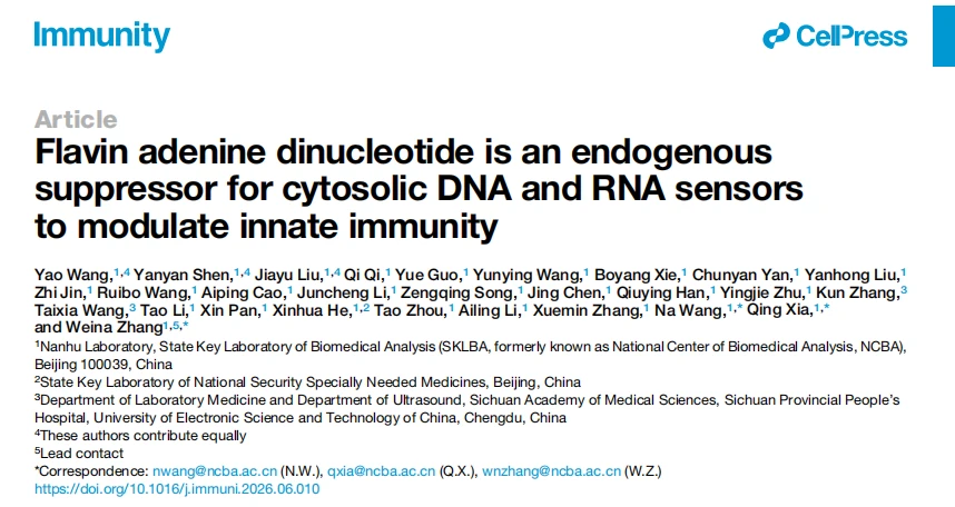
Immunity Study | Flavin Adenine Dinucleotide (FAD) Acts as a Dual Endogenous Inhibitor of cGAS and RIG-I Nucleic Acid Sensing

Introduction
The cytosolic nucleic acid sensors cGAS and RIG-I are key molecules that initiate type I interferon (IFN-I) innate immune responses. They recognize cytosolic double-stranded DNA and double-stranded RNA, respectively, to activate antiviral and antitumor immunity. However, persistent activation of nucleic acid-sensing pathways can lead to pathological conditions such as autoinflammation and cellular senescence. Current studies have largely focused on regulatory factors of cGAS or RIG-I individually, while endogenous regulatory molecules capable of simultaneously suppressing both major sensors have remained unidentified. The mechanisms by which metabolites regulate immune homeostasis therefore remain to be fully elucidated. Recently, a research team from Nanhu Laboratory published a study in Immunity aimed at elucidating the endogenous metabolic mechanisms that balance nucleic acid-sensing pathway activation and immune tolerance, revealing a novel pathway through which nutrient metabolism regulates innate immune homeostasis. The study used FLAD1 knockout A549 cells provided by Ubigene as a core experimental model , providing critical cellular support for establishing the complete “FLAD1-FAD-mediated inhibition of cGAS/RIG-I” causal pathway and validating the dual inhibitory function of FAD.
Workflow
- 1. Metabolite screening: An ISRE fluorescent reporter system was used to screen an endogenous metabolite library, identifying FAD as a potent inhibitor of nucleic acid-induced IFN-I responses. At the cellular level, FAD specifically blocked IFN-β secretion induced by polyI (RNA) and HT-DNA (DNA), without affecting TLR pathway activation.
- 2. Analysis of the upstream metabolic pathway: Viruses and cytosolic nucleic acids directly bound to FLAD1 and inhibited its enzymatic activity, thereby reducing cellular FAD levels. FLAD1 deficiency directly relieved FAD-mediated inhibition of cGAS/RIG-I and enhanced IFN responses.
- 3. Molecular interaction and structural mechanism: SPR, pull-down assays, and AlphaFold3 molecular docking demonstrated that FAD directly binds the catalytic domain of cGAS and the helicase domain of RIG-I, occupying substrate-binding pockets and competitively inhibiting enzymatic activity. Key amino acid residues involved in the interactions were also identified.
- 4. In vitro physiological validation: FLAD1 knockout and exogenous FAD rescue cell models were used to demonstrate that FAD deficiency enhances antiviral immunity, whereas FAD supplementation alleviates DNA damage-induced cellular senescence and SASP-associated inflammation.
- 5. In vivo animal phenotypes: Whole-body FLAD1 knockout mice and myeloid-specific FLAD1 knockout mice in a lupus model demonstrated that FAD deficiency enhances antiviral responses in vivo while exacerbating autoinflammation and lupus-associated tissue damage.
- 6. Clinical data validation: Analysis of the public GSE249619 dataset showed significantly reduced FLAD1 expression in monocytes from patients with systemic lupus erythematosus (SLE), which was associated with a high-IFN inflammatory phenotype.
Mechanistic Model
Key Findings
1. FAD Specifically Inhibits Cytosolic DNA/RNA-Induced IFN-I Signaling Without Affecting TLR Pathways
Cellular experiments showed that nucleic acid stimulation reduced FAD levels while increasing interferon production. Exogenous FAD dose-dependently inhibited cytosolic cGAS/RIG-I signaling without interfering with TLR signaling. In viral models, FAD pretreatment attenuated antiviral responses. Among the B vitamins tested, only vitamin B2 supported the synthesis of functional FAD, whereas the other B vitamins had no such effect. Inhibition of vitamin B2 uptake reduced FAD levels and enhanced interferon signaling, providing further evidence for the immunosuppressive role of FAD.
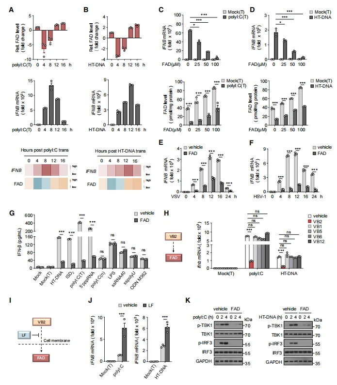
Figure 1. FAD specifically inhibits cytosolic nucleic acid-induced IFN-I signaling
2. Cytosolic Nucleic Acids Directly Inhibit FLAD1 Activity and Reduce FAD Synthesis, Relieving the Immune Brake
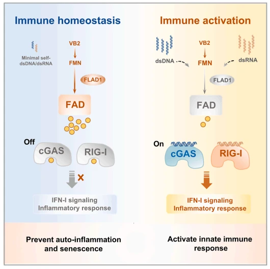
FLAD1 catalyzes the synthesis of FAD from FMN. Cytosolic double-stranded DNA and RNA directly bound to the N-terminal domain of FLAD1, noncompetitively inhibiting its enzymatic activity and reducing FAD production, whereas TLR ligands had no such effect. Nucleic acid stimulation progressively decreased intracellular FAD levels. FLAD1-knockout macrophages exhibited lower basal FAD levels and stronger interferon responses. Re-expression of wild-type FLAD1 or supplementation with exogenous FAD restored immune homeostasis, whereas an inactive FLAD1 mutant failed to rescue the phenotype.
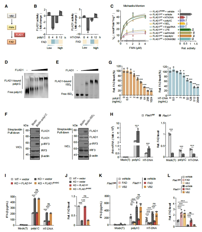
Figure 2. FLAD1 activity is directly inhibited by cytosolic nucleic acids, while FLAD1-associated FAD deficiency promotes activation of IFN-I signaling
3. FAD Directly Binds the Catalytic Pockets of cGAS and RIG-I to Competitively Block Their Enzymatic Activities
Biochemical and structural analyses demonstrated that FAD specifically binds cGAS and RIG-I, with binding affinities consistent with physiological FAD concentrations. FAD occupies the catalytic pockets of both sensors and competes with their substrates, with aromatic residues contributing to the interactions. The corresponding pockets in homologous proteins are too small to accommodate FAD. FAD specifically blocked the upstream enzymatic activities of cGAS and RIG-I without affecting activation of downstream signaling molecules such as STING and TBK1.
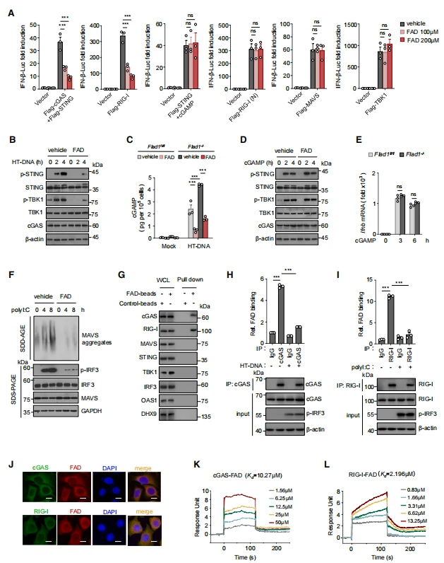
Figure 3. FAD targets nucleic acid sensors to limit IFN-I signaling
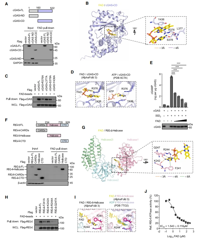
Figure 4. FAD binds to the catalytic pockets of nucleic acid sensors and inhibits their enzymatic activities
4. The FLAD1-FAD Axis Regulates Cellular Senescence and Sterile Inflammation
DNA-damaging agents, including doxorubicin (DOX), camptothecin (CPT), and H₂O₂, induced the release of endogenous nucleic acids into the cytosol, activating IFN responses and senescence-associated secretory phenotype (SASP) programs. Exogenous FAD treatment significantly reduced the expression of IFN- and SASP-associated genes and decreased the proportion of SA-β-Gal-positive senescent cells. Following DNA damage, Flad1⁻/⁻ mouse embryonic fibroblasts (MEFs) exhibited substantially aggravated senescence and inflammatory phenotypes, which were reversed by FAD supplementation. RNA-seq and GSEA further demonstrated significant enrichment of type I interferon, senescence, and inflammatory pathways in FLAD1-deficient cells, while FAD treatment reversed these pathway enrichments.
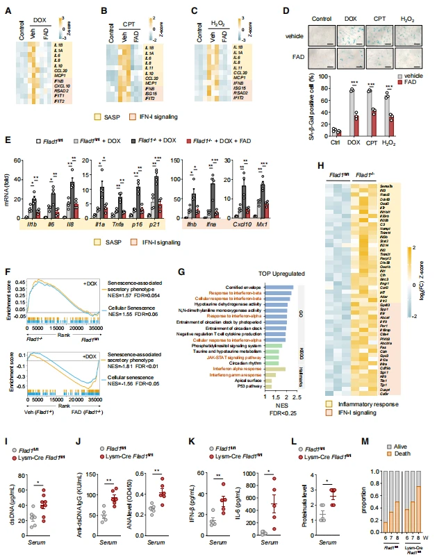
Figure 5. FLAD1-associated FAD suppresses autoinflammatory responses both in vitro and in vivo
5. In Vivo Studies: FAD Deficiency Enhances Antiviral Immunity but Exacerbates Autoimmune Lupus
Using antiviral models involving VSV and HSV infection, the study found that Flad1⁻/⁻ mice exhibited significantly reduced FAD levels in multiple tissues and serum. Following viral infection, serum IFN-β levels and tissue ISG expression were substantially higher than those in wild-type mice, while viral replication was significantly reduced. These mice also exhibited less inflammatory infiltration in the lungs and enhanced antiviral capacity. Primary macrophages isolated from Flad1⁻/⁻ mice showed markedly increased IFN secretion following viral stimulation, whereas in vitro FAD supplementation suppressed excessive interferon release.
In an imiquimod (IMQ)-induced lupus nephritis model, myeloid-specific Flad1 knockout mice (Lysm-Cre Flad1fl/fl) exhibited significantly elevated serum dsDNA, antinuclear antibodies (ANA), IFN-β, and IL-6 levels, accompanied by aggravated proteinuria, splenomegaly, and increased mortality. These findings indicate that FAD deficiency in myeloid cells leads to persistent activation of the cGAS/RIG-I pathways by endogenous nucleic acids, while chronically elevated IFN signaling promotes autoantibody production and renal injury.
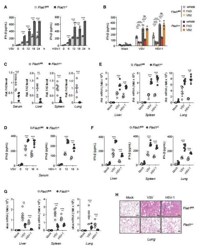
Figure 6. FLAD1-associated FAD deficiency enhances antiviral innate immune responses in vivo
Research Innovations
- Conceptual Innovation: This study is the first to identify a single endogenous metabolite, FAD, as a dual regulator capable of targeting both major cytosolic nucleic acid sensors, cGAS and RIG-I. It reveals a novel vitamin B2-FLAD1-FAD metabolic-immune regulatory axis and demonstrates how metabolic molecules maintain the balance between antiviral immunity and autoinflammatory homeostasis, addressing a previously uncharacterized mechanism for the coordinated regulation of nucleic acid-sensing pathways.
- Technical Innovation: By integrating SPR-based molecular affinity analysis, AlphaFold3 structural modeling, site-directed mutagenesis, and FLAD1 knockout cell rescue systems, the study established a comprehensive biochemical-cellular-structural research framework. This approach precisely elucidated how the small molecule interacts with the catalytic pockets of nucleic acid-sensing enzymes and provides a standardized research paradigm for the structural design of anti-inflammatory and antiviral small-molecule lead compounds.
Summary
This study demonstrates that FAD is an endogenous dual inhibitor of cGAS and RIG-I. Under homeostatic conditions, FAD suppresses both pathways to maintain immune tolerance. Accumulation of cytosolic nucleic acids inhibits FLAD1 and depletes FAD, thereby releasing antiviral immune responses. Cellular and animal experiments demonstrated that FAD deficiency enhances antiviral immunity but also promotes senescence and lupus-like autoinflammation, whereas FAD supplementation alleviates these pathological phenotypes. Clinical data further support the association between reduced FLAD1 expression and lupus. This study reveals a novel mechanism by which metabolism regulates nucleic acid sensing and provides a potential metabolic-targeting strategy for the treatment of infection, autoimmune disease, and inflammation associated with aging.
Support from Ubigene
The in vitro mechanistic studies in this work relied on FLAD1 Knockout Cell Line (A549) provided by Ubigene as a key experimental model. This model enabled the researchers to clearly establish the causal chain of “FLAD1 deficiency → decreased intracellular FAD → excessive activation of the cGAS/RIG-I pathways” and effectively exclude potential off-target interference caused by exogenous nucleic acids or small-molecule treatments. It therefore served as an essential experimental tool for establishing the complete FLAD1-FAD regulatory pathway.
Ubigene has always upheld the core philosophy of “Making Gene Editing Easier,” continuously iterating its products and services. To date, Ubigene has achieved 13,000+ successful gene editing cases and offers 10,000+ KO cell products. Based on its independently developed innovative technology, gene-editing efficiency can reach up to 97%. Ubigene has provided high-quality gene-editing services and products to more than 10,000 life science laboratories, pharmaceutical companies, and CROs worldwide.
If you are also interested in studying FLAD1, Ubigene offers multiple gene knockout cell models to support your research. Please contact us for more information.
 Promotions