
HepG2 Gene-Edited Cell Lines: Applications in Metabolic and Genetic Disease Research

HepG2 Cells: A Versatile Model for Liver Research
The HepG2 cell line was derived from the liver tissue of a 15-year-old Caucasian male patient with hepatocellular carcinoma [1] . Since its establishment in 1979, HepG2 has become a widely used cell model for liver biology, metabolic regulation, and drug development due to its retention of key liver-specific functions. Compared with primary hepatocytes, HepG2 cells have unlimited proliferative capacity , allowing stable long-term culture and passaging without the need for freshly isolated tissue. Their relatively stable genetic background also minimizes the inter-individual variation associated with primary cells, improving experimental reproducibility [2] . HepG2 cells also highly express liver-associated markers such as albumin, alpha-fetoprotein (AFP), and drug-metabolizing enzymes including members of the CYP450 family. They retain important physiological functions related to lipid metabolism and copper transport [2] . Compared with other hepatocellular carcinoma cell lines, such as Hep3B and SMMC-7721, HepG2 cells more closely resemble human hepatocytes in several functional characteristics. They are therefore widely used for studying liver metabolism, disease mechanisms, and drug screening.
Key Applications of HepG2 Cells
The characteristics of HepG2 cells make them useful for modeling a variety of liver-related physiological and pathological processes, particularly metabolic disorders and genetic diseases:
- Lipid Metabolism Disorder Models: HepG2 cells can be induced with high-fat culture conditions to model lipid accumulation associated with non-alcoholic fatty liver disease (NAFLD). Their triglyceride and cholesterol synthesis pathways are highly similar to those of human hepatocytes, making HepG2 a classic model for studying lipid metabolism and its regulation [1] .
- Drug Metabolism and Toxicity Assessment: HepG2 cells express key drug-metabolizing enzymes such as CYP3A4 and CYP2C9. They are therefore used to investigate drug metabolic pathways, metabolite toxicity, and drug interactions, providing important experimental data for drug development [1] .
- Copper Metabolism Disorder Models: HepG2 cells have functional pathways for copper uptake and export. By regulating the expression of copper transport proteins, HepG2 cells can be used to model disorders associated with abnormal copper metabolism, including Wilson disease [5] .
CRISPR/Cas9 Gene Editing in HepG2 Cells
Although HepG2 is an adherent cell line, conventional gene-editing approaches can be limited by low efficiency and off-target effects. With its precise targeting, simple design, and relatively low cytotoxicity, CRISPR/Cas9 provides an effective approach for investigating liver-related gene functions and constructing disease models in HepG2 cells.
LDLR Editing for Hypercholesterolemia Research
The low-density lipoprotein receptor (LDLR) is a key mediator of plasma LDL-C uptake. Defects in the LDLR gene can cause familial hypercholesterolemia (FH), which can lead to atherosclerosis and other cardiovascular diseases [3] . In one study, researchers analyzed 287 patients with familial hypercholesterolemia and 45 non-FH individuals. Exon-targeted sequencing identified six LDLR missense variants, and CRISPR/Cas9 was used to generate six HepG2 cell lines carrying these variants. The p.C184Y variant increased LDLR protein stability, while p.G373D and p.G549D moderately altered protein conformation. The p.V797L, p.R814Q, and p.V827I variants had relatively limited effects on protein stability. CRISPR/Cas9-edited HepG2 cells carrying p.G373D, p.V797L, or p.R814Q were further analyzed. All three variants significantly reduced LDLR expression and activity, resulting in abnormal lipid localization and substantial lipid accumulation in the cells. Clinical data showed that individuals carrying p.G549D, p.V797L, or p.R814Q had significantly elevated LDL cholesterol levels. The p.G373D and p.G549D variants were also closely associated with typical clinical manifestations of FH. Overall, the study demonstrated that different LDLR missense variants can alter protein structure and impair receptor function, contributing to abnormal lipid levels and the development and progression of familial hypercholesterolemia [3] . Other studies have shown that LDLR deficiency can reduce HBV infection efficiency by 80–90%. Restoring LDLR expression completely recovered the cells' susceptibility to HBV infection, while LDL overexpression further increased HBV infection. Experimental evidence indicated that LDLR does not affect HBV genome replication but mainly facilitates viral entry by binding apolipoprotein E on the viral envelope and acting as a cellular attachment receptor. These findings highlight the role of LDLR in HBV infection and provide a potential target and experimental basis for the development of new antiviral therapies [4] .
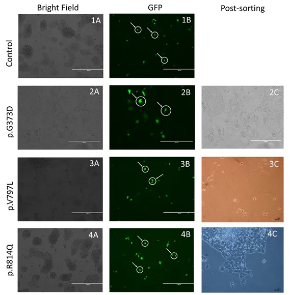
Figure 1. Flow cytometric sorting of HepG2 cells carrying LDLR missense variants
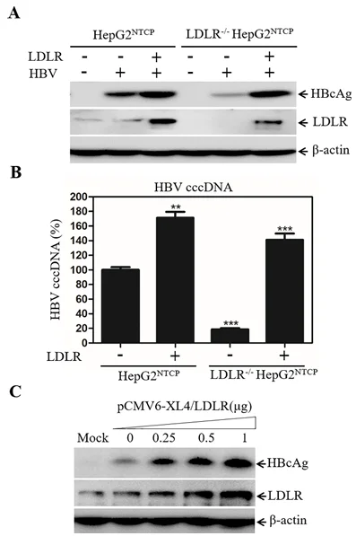
Figure 2. Increased HBV infection efficiency following LDLR restoration
ATP7B Editing for Wilson Disease Modeling
The ATP7B gene encodes a copper-transporting protein. Mutations or loss of ATP7B impair copper excretion through bile, resulting in abnormal copper accumulation in the liver and contributing to Wilson disease [5] . Researchers found that ATP7B can directly interact with the autophagy marker LC3B through its LIR3 motif. Copper stimulation induces autophagy in wild-type HepG2 cells. In contrast, ATP7B-knockout cells show impaired conversion of LC3B-I to LC3B-II after copper treatment, together with significantly reduced autophagosome–lysosome fusion. Restoring wild-type ATP7B expression in ATP7B-deficient cells effectively rescued copper-induced autophagy. However, an ATP7B protein carrying an LIR3-site mutation could not properly localize to the autophagosomal membrane and failed to restore the autophagy-deficient phenotype. These findings demonstrate that the interaction between ATP7B and LC3B plays an important role in coordinating copper metabolism and autophagy to eliminate excess intracellular copper. This provides important experimental evidence for understanding the mechanisms underlying Wilson disease and developing targeted interventions [5] . This model provides a useful tool for investigating the pathogenesis of Wilson disease and screening strategies to mitigate copper toxicity, supporting the development of disease-specific therapies [6] .
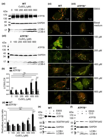
Figure 3. Effects of ATP7B knockout on copper-induced autophagy in HepG2 cells
Efficient HepG2 Gene Editing with EZ-editor™
EZ-editor™ is Ubigene's proprietary technology for cell line gene editing based on CRISPR/Cas. It is designed for the efficient generation of gene knockout cell lines. The technology integrates multiple capabilities, including a proprietary algorithm for designing gRNAs based on cell-specific genomic characteristics, methods for optimizing gene-editing parameters across thousands of cell lines, precise approaches for evaluating Cell Pool editing efficiency, strategies for improving monoclonal formation rates, and high-throughput methods for genotyping cells from limited samples. Compared with conventional approaches, EZ-editor™ can increase gene-editing efficiency by 10–20-fold.
Using EZ-editor™ technology, Ubigene has successfully generated multiple gene knockout HepG2 cell lines targeting genes of interest in metabolic and disease research. Inquire now!
References
- [1]Knowles BB, Howe CC, Aden DP. Human hepatocellular carcinoma cell lines secrete the major plasma proteins and hepatitis B surface antigen. Science. 1980 Jul 25;209(4455):497-9. doi: 10.1126/science.6248960. PMID: 6248960.
- [2]Busch SJ, Barnhart RL, Martin GA, Flanagan MA, Jackson RL. Differential regulation of hepatic triglyceride lipase and 3-hydroxy-3-methylglutaryl-CoA reductase gene expression in a human hepatoma cell line, HepG2. J Biol Chem. 1990 Dec 25;265(36):22474-9. PMID: 2176219.
- [3]Mori AA, Malaquias VB, Bonjour K, Ferreira GM, Bortolin RH, Borges JB, Oliveira VF, Gonçalves RM, Faludi AA, Bastos GM, Thurow H, Sampaio MF, Ciconelli RM, Cury AN, Fajardo CM, Hirata RDC, Hirata MH. Effects of LDLR variants rs5928, rs750518671 and rs879254797 on protein structure and functional activity in HepG2 cells transfected with CRISPR/Cas9 constructs. Gene. 2024 Jan 10;890:147821. doi: 10.1016/j.gene.2023.147821. Epub 2023 Sep 20. PMID: 37739193.
- [4]Dullens SP, Mensink RP, Bragt MC, Kies AK, Plat J. Effects of emulsified policosanols with different chain lengths on cholesterol metabolism in heterozygous LDL receptor-deficient mice. J Lipid Res. 2008 Apr;49(4):790-6. doi: 10.1194/jlr.M700497-JLR200. Epub 2007 Dec 27. PMID: 18162663.
- [5]Pantoom S, Pomorski A, Huth K, Hund C, Petters J, Krężel A, Hermann A, Lukas J. Direct Interaction of ATP7B and LC3B Proteins Suggests a Cooperative Role of Copper Transportation and Autophagy. Cells. 2021 Nov 10;10(11):3118. doi: 10.3390/cells10113118. PMID: 34831341; PMCID: PMC8625360.
- [6]Yamaguchi Y, Heiny ME, Suzuki M, Gitlin JD. Biochemical characterization and intracellular localization of the Menkes disease protein. Proc Natl Acad Sci U S A. 1996 Nov 26;93(24):14030-5. doi: 10.1073/pnas.93.24.14030. PMID: 8943055; PMCID: PMC19489.
 Promotions