
A Comprehensive Guide to ID8-Luc Cells: A powerful Tool for Ovarian Cancer Research


Literature Overview
In ovarian cancer research, the ability to dynamically and visually track tumor growth, progression, and therapeutic response has long been a critical challenge in in vivo studies. Traditional animal experiments rely on tumor volume measurements, body weight, abdominal girth, and endpoint histological analyses. However, for ovarian cancer, which is a disease characterized by widespread peritoneal dissemination and malignant ascites formation, endpoint indicators alone often fail to capture the continuous disease course.
ID8-Luc cells offer a valuable research model to address this challenge. By stably expressing firefly luciferase in murine ovarian cancer ID8 cells, researchers can use bioluminescence imaging (BLI/IVIS) to dynamically and non-invasively monitor tumor burden, converting invisible tumor progression into quantifiable luminescent signals. In recent years, ID8-Luc cells have been widely used in studies of ovarian cancer development and progression, the tumor microenvironment, drug evaluation, immunotherapy, and combination therapies.
Applications of ID8-Luc Cells
ID8-Luc cells are widely used to establish syngeneic murine models of intraperitoneal ovarian cancer*, Following intraperitoneal inoculation, these cells recapitulate key features of human ovarian cancer progression, including the formation of multiple peritoneal tumor nodules and malignant ascites, thereby closely modeling the in vivo microenvironment of advanced epithelial ovarian cancer. Importantly, these models retain an intact mouse immune system, overcoming a major limitation of human tumor xenograft models, including patient-derived xenografts (PDXs), which are generally immunodeficient.
(*The model uses C57BL/6-derived ID8-Luc ovarian cancer cells for intraperitoneal inoculation into immunocompetent C57BL/6 mice with a fully matched genetic background, thereby modeling key features of advanced human epithelial ovarian cancer.)
Major Research Applications:
- Immunotherapy Drug Evaluation: ID8-Luc cells can be used for in vivo efficacy evaluation of antibodies, antibody-drug conjugates (ADCs), immune checkpoint inhibitors, and macrophage-targeted therapeutics. With the aid of non-invasive, longitudinal bioluminescence imaging (IVIS), researchers can dynamically monitor tumor burden, intraperitoneal metastasis, and ascites progression without sacrificing the animals, enabling long-term assessment of treatment response and changes in survival [1] .
- Tumor Microenvironment Studies: Ascites and intraperitoneal immune cells can be collected to characterize immune populations, including tumor-associated macrophages (TAMs), myeloid-derived suppressor cells (MDSCs), regulatory T cells (Tregs), and CD8⁺T cells. These analyses can be used to investigate immunosuppressive tumor microenvironments and the mechanisms underlying resistance to immunotherapy [2] .
- Development of Combination Therapies: ID8-Luc models can be used to evaluate the synergistic effects of immunotherapy in combination with chemotherapy, radiotherapy, or antibody-drug conjugates. They can also support mechanistic investigations and ex vivo functional assays related to intraperitoneal phagocytosis and antigen presentation [4] .
- Tumor Metastasis and Immune Memory Studies: These models can be used to investigate intraperitoneal dissemination and metastasis, as well as to evaluate the development of antitumor immune memory following treatment [3] .
Case Studies of ID8-Luc Cells Application in Research
1. Application of ID8-Luc Cells in Preclinical Studies of CD24-Targeted Macrophage Immunotherapy
Journal |
Clinical Cancer Research
(IF=10.9)
Title |
Targeting CD24 Activates Macrophages to Reduce Tumor Burden in
Preclinical Models of Solid Tumors
This study developed PHST001, a humanized IgG4 anti-CD24 antibody that blocks the CD24-SIGLEC-10 pathway, activating macrophage-mediated tumor cell phagocytosis. The anti-tumor efficacy as monotherapy and in combination with radiotherapy, chemotherapy, or ADCs was validated across multiple solid tumor models. Mechanistically, tissue-resident TIM4⁺ macrophages were identified as the primary effector cells, and upon tumor phagocytosis, these macrophages mediated antigen cross-presentation, activating CD8⁺ T cells and establishing anti-tumor immunological memory. The authors also note that a Phase I clinical trial (NCT06840886) is underway [1] .

Role of ID8-Luc Cells in This Study
Parental ID8-Luc cells (provided by Ubigene, Catalog# YC-C103-Luc-P)
were engineered to stably express human CD24 through lentiviral
overexpression, generating ID8-huCD24-Luc stable cells. These cells were
intraperitoneally implanted into C57BL/6 mice for ex-vivo peritoneal
phagocytosis assays, directly demonstrating that PHST001 preferentially
activates TIM4⁺ tissue-resident macrophages to engulf tumor cells.
Additionally, they were used for in vivo monitoring of PHST001-mediated
suppression of peritoneal tumors and ascites, and for analyzing changes
in CD8⁺ T and Treg immune subsets within the peritoneal cavity,
confirming that CD24 blockade bridges innate and adaptive anti-tumor
immunity.
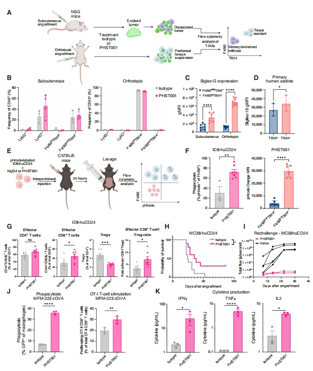
Figure 1. PHST001 activates tissue-resident macrophages and CD8⁺ T cells
2. Application of ID8-Luc in Preclinical Research on a PD-L1-Targeted pADC (U6244-051)
Journal |
Journal of Controlled Release
(IF=11.5)
Title |
PDL1 Targeted AntibodyPolymerEpirubicin Conjugate Prolongs Survival in a
Preclinical Murine Model of Advanced Ovarian Cancer
The authors constructed U6244-051, a conjugate with a DAR of 42-43 that combines PD-L1 blockade with chemotherapeutic payload release. In vitro, U6244-051 induced immunogenic cell death and increased the expression of calreticulin and MHC class I. In an advanced ID8-Luc syngeneic ovarian cancer model with malignant ascites, treatment achieved 100% survival at 100 days, even when therapy was initiated at an advanced stage of disease. Furthermore, it remodeled the ascites immune microenvironment by reducing TAMs, MDSCs, and Tregs while increasing activated CD8⁺ T cells, with repeated dosing amplifying these immunomodulatory effects [3] .
Role of ID8-Luc Cells in This Study
As the core cellular model in this study, ID8-Luc cells (provided by
Ubigene, Catalog# YC-C103-Luc-P) were inoculated intraperitoneally to
establish an advanced ovarian cancer model characterized by malignant
ascites. Bioluminescence imaging was used to dynamically monitor tumor
burden and evaluate anti-tumor efficacy. Ascites were isolated for flow
cytometric analysis to systematically profile the immune landscape of
early- and late-stage ovarian cancer ascites, compare immune cell
dynamics across different dosing regimens, assess the pADC's remodeling
effects on the tumor microenvironment, and validate the
immuno-chemotherapeutic synergy of the conjugate.
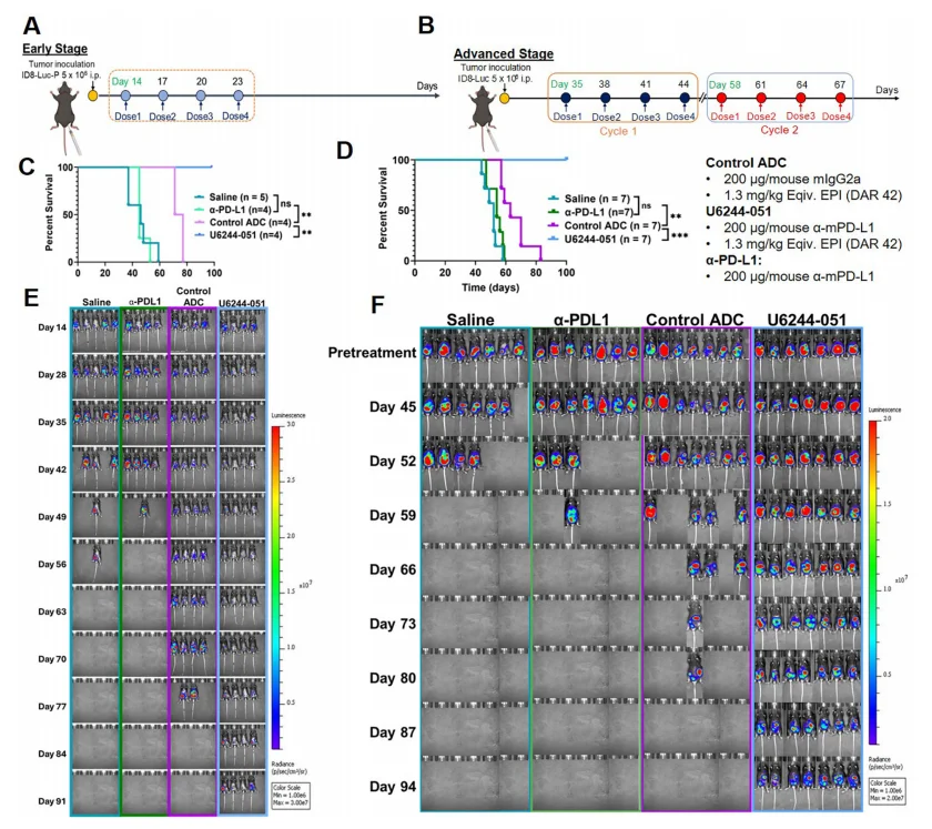
Figure 2. U6244-051 significantly prolongs survival and reduces tumor burden in ID8-Luc tumor-bearing mice
Conclusion
With their syngeneic, immunocompetent characteristics and their ability to closely recapitulate key features of advanced ovarian cancer, including malignant ascites and intraperitoneal dissemination, ID8-Luc cells are widely used in preclinical studies of ovarian cancer immunotherapy, combination therapies, tumor microenvironment, immune memory, and macrophage-targeted therapies. The model can be used not only for in vivo pharmacodynamic and efficacy evaluation, but also for in vitro cellular functional assays and ex vivo intraperitoneal functional studies. As such, ID8-Luc cells serve as an important tool for bridging molecular mechanisms with in vivo therapeutic efficacy and provide a reliable preclinical platform for the translational development of innovative therapies, including antibodies, ADCs, and nanomedicines.
Ubigene Luciferase Stable Cell Lines
Ubigene currently offers more than 300 Luc stable cell lines, all generated using lentiviral transduction and engineered to stably express firefly luciferase (Firefly Luciferase, Luc). All cell lines have undergone luciferase activity testing and feature high signal specificity, high sensitivity, stable imaging performance, and quantifiable bioluminescence signals. If you are also interested in using Luc-expressing cells for in vivo pharmacodynamic evaluation, tumor metastasis studies, or related research, please feel free to contact us for more information!
Reference
[1]Faget DV, Brewer RE, Sampaio J, Blacker G, Forcina GC, Qazi S,
Malusare PR, Ludwig S, Hart KE, Hansen JM, Beans A, Virani R, Dorigo O,
Rousseau R, Ko PJ, Cao JY, Burg JS, Majeti R, Weissman IL, Barkal AA,
Maute RL, Kahn SA. Targeting CD24 Activates Macrophages to Reduce Tumor
Burden in Preclinical Models of Solid Tumors. Clin Cancer Res. 2026 Sep
1;32(17):3934-3955. doi: 10.1158/1078-0432.CCR-26-0481. PMID: 42478960;
PMCID: PMC13530989.
[2]Hamze Sinno S, Imperatore JA, Bai S, Gomes-Jourdan N, Mafarachisi N,
Coronnello C, Zhang L, Jašarević E, Osmanbeyoglu HU, Buckanovich RJ,
Cascio S. Egfl6 promotes ovarian cancer progression by enhancing the
immunosuppressive functions of tumor-associated myeloid cells. J Clin
Invest. 2024 Nov 1;134(21):e175147. doi: 10.1172/JCI175147. PMID:
39312740; PMCID: PMC11527450.
[3]Li J, Al Faruque H, Li S, Sima M, Sborov D, Hu-Lieskovan S, Werner T,
Kopeček J, Yang J. PD-L1 targeted antibody-polymer-Epirubicin conjugate
prolongs survival in a preclinical murine model of advanced ovarian
cancer. J Control Release. 2025 Jun 10;382:113682. doi:
10.1016/j.jconrel.2025.113682. Epub 2025 Apr 2. PMID: 40185333; PMCID:
PMC12912222.
[4]Adams SF, Grimm AJ, Chiang CL, Mookerjee A, Flies D, Jean S, McCann
GA, Michaux J, Pak H, Huber F, Neal C, Dangaj D, Bassani-Sternberg M,
Rusakiewicz S, Facciabene A, Coukos G, Gimotty PA, Kandalaft LE. Rapid
tumor vaccine using Toll-like receptor-activated ovarian cancer ascites
monocytes. J Immunother Cancer. 2020 Aug;8(2):e000875. doi:
10.1136/jitc-2020-000875. PMID: 32817208; PMCID: PMC7430560.
 Promotions