Nanogel Stiffness Reprograms TAMs and Suppresses Tumor Growth

Introduction
Tumor-associated macrophages (TAMs) predominantly exhibit an anti-inflammatory, tumor-promoting phenotype and are a key component of the immunosuppressive tumor microenvironment. Macrophages can be reprogrammed by macroscale mechanical stimuli, but whether the mechanical properties of individual nanoparticles can regulate TAM function has remained unclear.
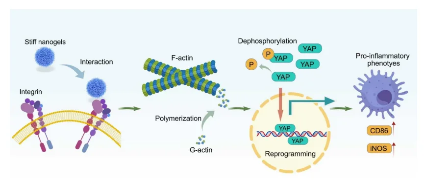
The research team showed that adjusting the crosslinking density of nanogels changed their stiffness. Stiff nanogels (15% NGs, Young’s modulus of 551 kPa) reprogrammed anti-inflammatory macrophages toward a pro-inflammatory phenotype more effectively than soft nanogels (2% NGs, 65 kPa). The stiffer formulation interacted more strongly with the β2 integrin subunit ITGB2, triggering actin polymerization and promoting YAP1 dephosphorylation and nuclear translocation. These changes increased CD86 and iNOS expression and stimulated TNF-α secretion, leading to tumor cell apoptosis.
The 15% NGs accumulated preferentially in the liver and significantly suppressed tumor growth in an orthotopic H22 liver cancer model. Their effects were also evaluated in patient-derived hepatocellular carcinoma (HCC) tissue explants. These findings highlight nanogel stiffness as a design parameter for cancer nanomedicines. Ubigene supplied H22-Luc cells (#YC-C053-Luc-P) for this study, providing a key in vivo tracking tool for establishing the orthotopic liver cancer model and monitoring therapeutic response over time.
Experimental Workflow
- Nanogel synthesis and phenotypic screening: The researchers synthesized nanogels of different stiffnesses and found that the 15% NGs were more effective at shifting anti-inflammatory macrophages toward a pro-inflammatory phenotype.
- Identification of the downstream effector: Measurements of YAP1 expression, phosphorylation, and nuclear translocation, together with verteporfin inhibition, identified YAP1 as a key downstream effector.
- Mapping of the upstream pathway: F-actin measurements, Lat-A inhibition, and ITGB2 neutralization established the sequence from ITGB2 engagement to actin polymerization and YAP1 activation.
- Functional validation in animal models: Transcriptomic and TNF-α pathway analyses, subcutaneous 4T1 tumor experiments, and intravenous treatment in the orthotopic H22 model demonstrated tumor suppression and preferential liver accumulation of the stiff formulation.
- Evaluation in human-derived models: Experiments with THP-1-derived macrophages and organotypic slices of patient-derived HCC tissue evaluated macrophage reprogramming and tumor cell apoptosis in human systems.
Proposed Mechanism
Research Background
The physicochemical properties of nanomedicines, including size, shape, and surface chemistry, substantially affect delivery to tumors. Research has also shown that nanoparticle mechanics can influence tumor accumulation and cellular uptake. Macrophages express mechanosensitive proteins on their surface, including integrins and Piezo1, which convert extracellular mechanical cues into biochemical signals. Inside the cell, the mechanosensitive regulator YAP1 links mechanical cues to changes in gene expression. Previous studies have shown that macroscale stimuli, such as matrix stiffness and stretching, can reprogram macrophages through pathways involving CD11b, Piezo1, and YAP/TAZ. How macrophages sense and respond to mechanical cues from individual nanoparticles remains less well understood.
Most TAMs exhibit an anti-inflammatory, tumor-promoting phenotype that contributes to immune evasion and tumor progression. Reprogramming these macrophages toward a pro-inflammatory phenotype is a potential immunotherapy strategy. The team had previously found that altering nanogel stiffness could reduce clathrin transcription and inhibit endocytosis in RAW264.7 macrophages. This led the researchers to investigate whether nanoparticle mechanics could also regulate TAM reprogramming.
Key Findings
1. Stiff Nanogels More Effectively Reprogram Macrophages
The researchers synthesized two P(NIPMAM-ss-MAA) nanogel formulations with similar particle sizes of approximately 220 nm and different stiffnesses. The soft 2% NGs had a Young’s modulus of 65 kPa, and the stiff 15% NGs had a Young’s modulus of 551 kPa. RAW264.7 macrophages were first polarized toward an anti-inflammatory phenotype with IL-4 and IL-13, then incubated with either formulation. The 15% NGs more effectively shifted the cells toward a pro-inflammatory phenotype. They significantly increased CD86 and iNOS at both the mRNA and protein levels and increased nitric oxide production. The anti-inflammatory markers CD206 and Arg-1 showed no appreciable change. Similar results were observed in bone marrow-derived macrophages (BMDMs). Although cells internalized the 2% NGs more efficiently, the 15% NGs induced stronger reprogramming. This pattern points to stiffness as the main driver of the observed response.
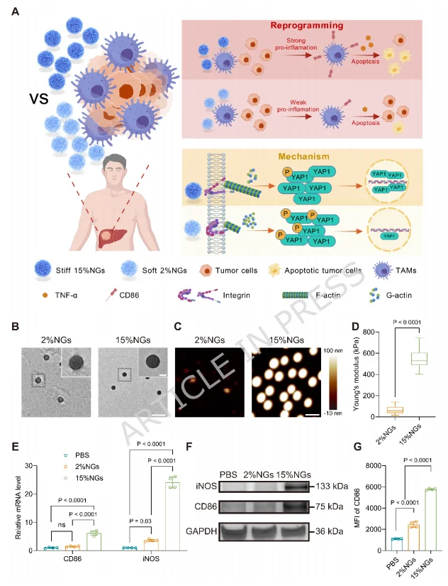
Figure 1. Effects of nanogels with different stiffnesses on macrophage reprogramming.
2. YAP1 Signaling Is Required for Nanogel-Induced Reprogramming
The 15% NGs significantly increased YAP1 expression, reduced its phosphorylation, and promoted its nuclear translocation. Verteporfin (VP), a YAP inhibitor, blocked the increase in iNOS and CD86 induced by the 15% NGs, supporting a requirement for YAP1 signaling in this response.
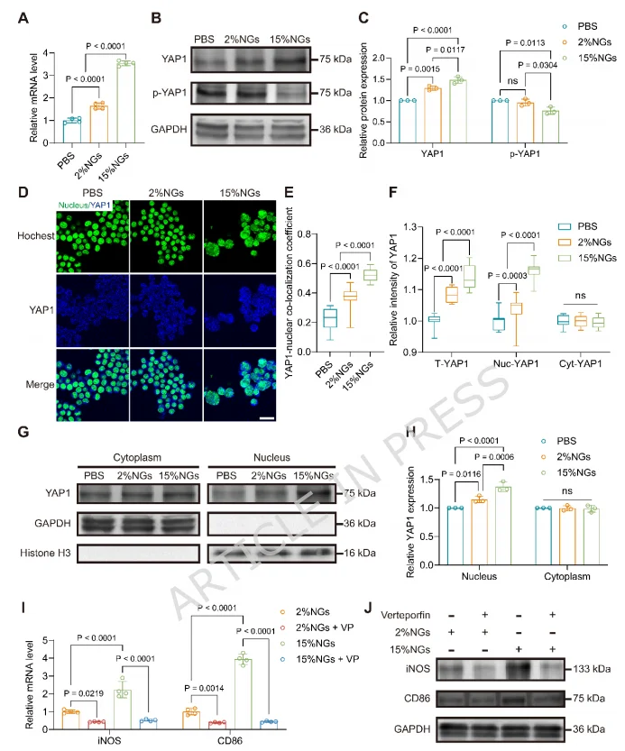
Figure 2. YAP1 signaling is required for nanogel-induced reprogramming of RAW264.7 macrophages toward a pro-inflammatory phenotype.
3. Stiff Nanogels Activate YAP1 Through Actin Polymerization
The 15% NGs significantly promoted actin polymerization, as indicated by an increase in the ratio of filamentous actin (F-actin) to globular actin (G-actin). Latrunculin A (Lat-A), an inhibitor of actin polymerization, attenuated the nanogel-induced increase in YAP1 expression, inhibited YAP1 dephosphorylation and nuclear translocation, and blocked the upregulation of pro-inflammatory markers. These results place actin polymerization upstream of YAP1 in the mechanical signaling pathway.
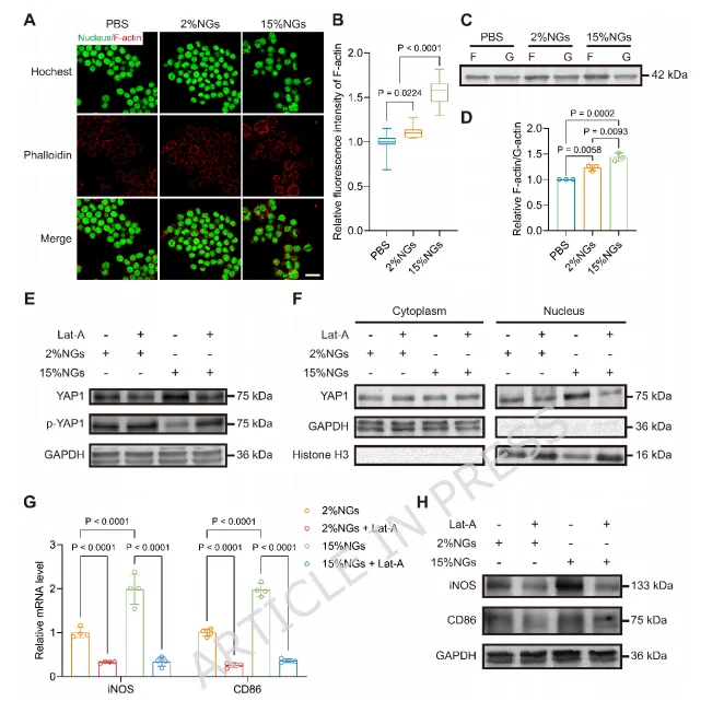
Figure 3. Stiff nanogels activate YAP1 signaling through actin polymerization.
4. Integrin Binding Initiates Pro-Inflammatory Reprogramming
The 15% NGs upregulated ITGB2, the β2 integrin subunit. Co-immunoprecipitation showed an increased association between ITGB2 and actin, while fluorescence colocalization and native PAGE indicated stronger binding between the 15% NGs and integrin αMβ2. An ITGB2-neutralizing antibody (anti-CD18) blocked actin polymerization, YAP1 activation, and pro-inflammatory reprogramming. Together, these experiments identified a signaling pathway linking integrin engagement to actin polymerization and YAP1 activation.
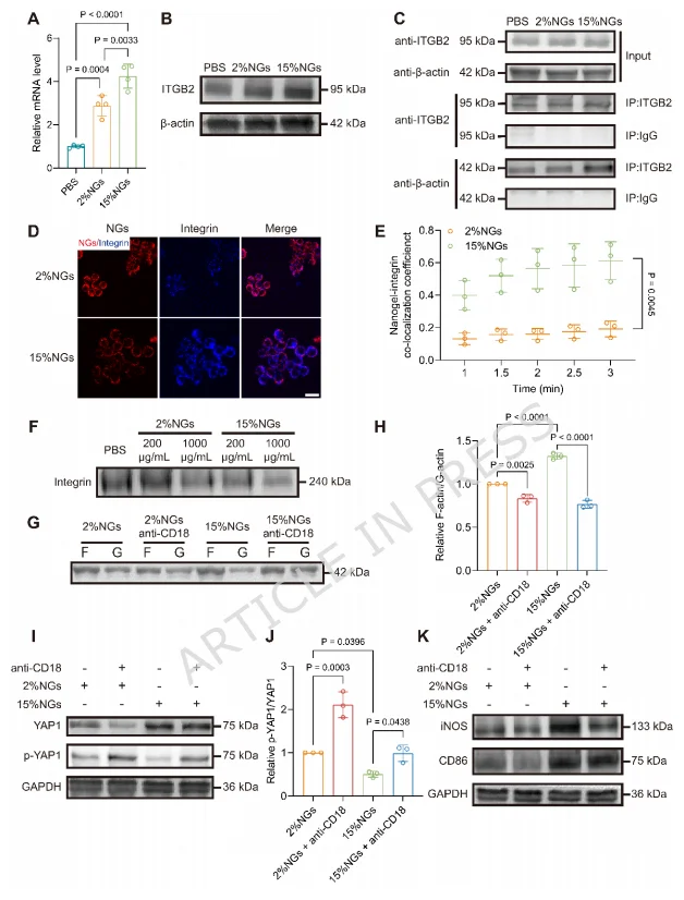
Figure 4. Nanogels of different stiffnesses initiate pro-inflammatory reprogramming through interactions with integrins.
5. Stiff Nanogels Suppress 4T1 Tumor Growth Through Macrophage TNF Signaling
Transcriptomic analysis identified 845 differentially expressed genes between the 15% and 2% NG treatment groups. KEGG analysis showed enrichment in several inflammatory pathways, and gene set enrichment analysis (GSEA) indicated significant activation of TNF signaling. The 15% NGs increased TNF-α transcription and secretion in macrophages. Conditioned medium from these macrophages significantly reduced 4T1 cell viability and induced apoptosis. Etanercept, which binds TNF-α and blocks its interaction with cell-surface receptors, reversed this effect. In a subcutaneous co-inoculation model, macrophages treated with 15% NGs suppressed tumor growth to a degree comparable to M1 macrophages and were significantly more effective than macrophages treated with 2% NGs. Intratumoral injection experiments also showed that the 15% NGs could reprogram TAMs in situ.
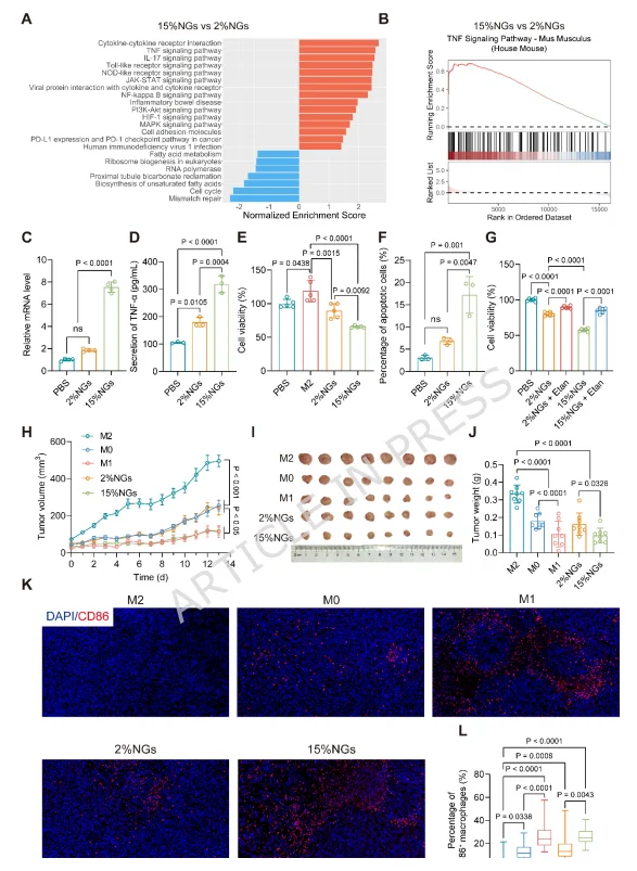
Figure 5. Stiff nanogels suppress 4T1 tumor growth by activating TNF signaling in macrophages.
6. Stiff Nanogels Suppress Orthotopic Liver Tumor Growth Through Tumor Accumulation and TAM Reprogramming
The 15% NGs accumulated more extensively in the liver and at tumor sites than the 2% NGs. In vitro, the stiff formulation increased macrophage cytotoxicity against H22 cells and enhanced their phagocytosis of these cells. The 15% NGs also significantly suppressed tumor growth in a subcutaneous H22 co-inoculation model. In the orthotopic H22 liver cancer model, bioluminescence imaging showed that intravenous administration of 15% NGs suppressed tumor growth. Tumor volume and weight were significantly reduced, and CD86 expression increased in TAMs. Blood biochemistry and hematoxylin and eosin (H&E) staining of major organs indicated good tolerability under the conditions tested.
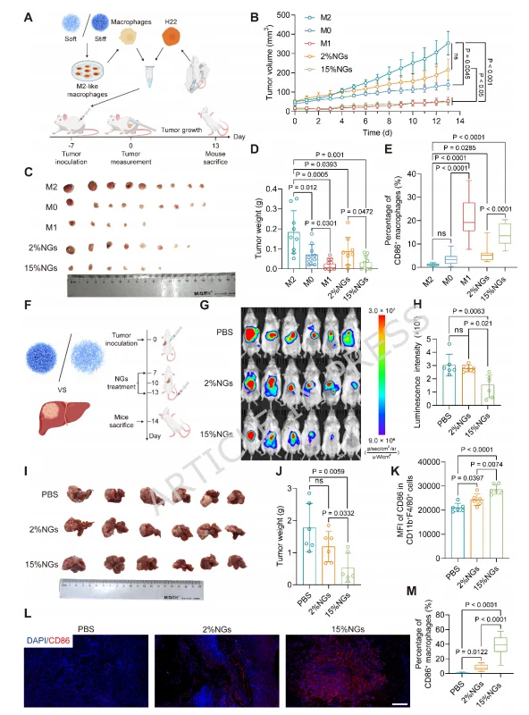
Figure 6. Stiff nanogels suppress orthotopic liver tumor growth through tumor accumulation and reprogramming of tumor-associated macrophages.
7. Stiff Nanogels Reprogram TAMs and Induce Tumor Cell Apoptosis in Patient-Derived HCC Tissue
Analysis of the TIMER 2.0 database showed that ITGB2 and YAP1 expression correlated positively with M1 macrophage infiltration in liver cancer. Higher expression was also associated with longer progression-free survival. In human THP-1-derived macrophages, the 15% NGs activated the ITGB2–actin–YAP1 pathway, shifted macrophages toward a pro-inflammatory phenotype, and enhanced their inhibitory effects on HepG2 and Huh7 cells. In organotypic HCC tissue slices from two patients, treatment with 15% NGs significantly increased CD86 expression and TNF-α production in TAMs and induced tumor cell apoptosis. These results support further investigation in human-derived models.
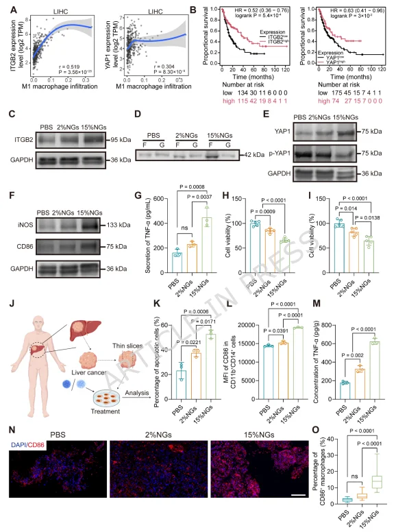
Figure 7. Stiff nanogels reprogram tumor-associated macrophages toward a pro-inflammatory phenotype and induce tumor cell apoptosis in patient-derived HCC tissue.
Summary
Nanogel stiffness acted as a nanoscale mechanical cue. The stiff formulation engaged ITGB2, promoted actin polymerization, and induced YAP1 dephosphorylation and nuclear translocation. This signaling pathway shifted TAMs toward a pro-inflammatory phenotype, increased TNF-α secretion, and induced tumor cell apoptosis. The 15% NGs bound integrins more strongly and showed greater liver accumulation. They significantly suppressed tumor growth in an orthotopic liver cancer model, and their effects on TAM reprogramming and tumor cell apoptosis were also observed in patient-derived HCC explants. The study maps a pathway through which nanoparticle mechanics can reshape TAM behavior and provides an experimental framework for incorporating stiffness into cancer nanomedicine design.
How Ubigene Supported the Study
Ubigene supplied H22-Luc cells (#YC-C053-Luc-P) to establish the
orthotopic liver cancer model and track tumor response over time. The
luciferase reporter enabled in vivo bioluminescence imaging, providing a
visual and quantifiable measure of tumor growth during treatment with
stiff nanogels.
Ubigene is committed to making gene editing easier and continually
improving its products and services. We have completed more than 13,000
successful gene editing projects and offer over 10,000 knockout cell
products. Our proprietary technologies achieve gene editing efficiencies
of up to 97%. Ubigene has provided gene editing products and services to
more than 10,000 life science laboratories, pharmaceutical companies,
and CROs.
If you are planning research with related tumor models, we offer a
range of research cell lines and stable cell lines.
Contact us to learn more!
 Promotions