Comprehensive Guide to Point Mutation Cell Line Construction via RNP Method


In today's era of increasingly popular high-throughput sequencing, a large number of human single nucleotide polymorphisms (SNPs) have been discovered to be closely related to various diseases. To reveal the specific roles of these SNPs in pathological processes, researchers increasingly rely on cell models with site-specific mutations as essential tools for functional research and mechanistic exploration. However, compared to gene knockout cell lines, the construction of point mutation cell lines involves complex molecular cloning techniques and high-throughput screening. Editing efficiency varies significantly across different cell lines, making the construction relatively difficult. Today, we will systematically review the methods and principles of constructing point mutation cell lines using the RNP method , and the factors that determine success.
I. Application Value of Point Mutation Cell Lines: Which Fields Are Inseparable from It?
A Point Mutation Cell Line refers to a cell model in which one or several base changes (point mutations) are introduced at a specific locus in the cell genome. It has become an essential tool for studying protein function, signaling pathways, disease modeling, and drug target validation. Whether for SNP correction, amino acid substitution, promoter mutation, or pathogenic mutation modeling, point mutation is one of the most common types of gene editing.
(I) Disease Mechanism Research
Point mutation cell models can accurately simulate the molecular and phenotypic changes triggered by specific mutations of specific genes at precise loci, making them particularly suitable for studying monogenic diseases and tumor driver mutations. For example:
- Tumor Research: Cell models with oncogenic driver mutations such as EGFR T790M and KRAS G12D are widely used to dissect the mechanisms of tumorigenesis and screen targeted drugs.
- Genetic Disease Modeling: Disease-related SNV cell models, such as TP53 R175H and HBB E6V (sickle cell disease), provide pivotal tools for studying the pathological mechanisms of hereditary diseases.
- Immunodeficiency Diseases: RAG1 gene point mutation cell lines can be used to study the molecular mechanisms of immune cell development defects. Researchers utilized the optimized Cas9 RNP technology to successfully construct a pre-B lymphocyte line with a homozygous RAG1 c.946T>G point mutation. They discovered that this mutation led to the downregulation of RAG1 and RAG2 protein expression and an increased proportion of cell apoptosis, providing an experimental basis for establishing single-nucleotide mutation disease models.
(II) Drug Screening and Target Validation
Accurate point mutation models can be utilized to validate the efficacy of candidate drugs against specific mutations. For instance, performing targeted drug screening in cell lines harboring KRAS G12D, G12C, or EGFR L858R mutations allows for the evaluation of compound specificity and resistance mechanisms, thereby facilitating the development of personalized therapeutic strategies.
(III) Protein Function and Regulation Research
By mutating specific sites within a gene of interest into different amino acids (such as alanine scanning mutagenesis) or introducing phosphorylation-mimicking mutations, the structural-functional relationships of proteins can be meticulously dissected. Concurrently, introducing site-specific mutations into regulatory elements within intracellular signaling pathways (such as miRNA target sites and splicing regulatory sequences) allows for the study of impacts at the gene expression level. Compared to gene knockouts, point mutation models provide phenotypic information with a higher degree of precision in functional studies.
II. Core Principles of Constructing Point Mutation Cell Lines via the RNP Method
(I) Basic Principle: Cas9 RNP + HDR
The theoretical foundation for constructing point mutation cell lines is Homology-Directed Repair (HDR). The core workflow of the RNP method involves pre-incubating gRNA and Cas9 protein in vitro to form an RNP complex, which is then co-transfected into target cells along with a single-stranded oligonucleotide (ssODN) serving as the homologous template. The specific mechanism is as follows: guided by the gRNA, the Cas9 nuclease recognizes and cleaves the target DNA site, generating a double-strand break (DSB). Concurrently, two major DNA repair pathways exist within the cell: Non-Homologous End Joining (NHEJ) and Homology-Directed Repair (HDR). NHEJ can occur during almost any phase of the cell cycle and frequently results in random insertions or deletions (indels). Conversely, HDR primarily occurs during the S and G2/M phases and can precisely "write" the target mutation into the genome utilizing the introduced ssODN template. Therefore, the core challenge in constructing point mutations lies in: how to prompt cells to preferentially select the HDR repair pathway over the NHEJ pathway.
(II) Key Strategies to Improve Homology Directed Repair Efficiency
- Cell Cycle Synchronization: Since HDR primarily occurs during the S and G2 phases, maximizing the proportion of cells in these stages during transfection is a direct means to enhance HDR efficiency. Studies have demonstrated that utilizing hydroxyurea for cell cycle synchronization can boost HDR efficiency by 1.57-fold.
- Optimizing the Molar Ratio of Cas9 to gRNA: The ratio of Cas9 to gRNA directly influences cleavage efficiency and HDR outcomes. Research indicates that the integration efficiency of CRISPR/Cas9 is maximized under an equimolar ratio (1:1) or with a slight excess of Cas9. Furthermore, the distance between the single-stranded DNA donor and the target locus is a critical determinant of HDR efficiency; the closer the cleavage site is to the mutation site, the higher the HDR efficiency.
- Utilization of NHEJ Inhibitors: Indirect promotion of HDR can be achieved by pharmacologically inhibiting the NHEJ pathway. Studies show that the DNA-PKcs inhibitor Nu7441 significantly enhances HDR-mediated gene correction efficiency, achieving over a 10-fold increase in HDR efficiency in HeLa cells, reaching up to 53%. Nu7441 expands the opportunity window for HDR by modulating the cell cycle, which reduces the proportion of cells in the G1 phase and extends the S and G2/M phases.
- Optimizing Donor DNA Design: For point mutation construction, ssODN is a commonly used donor template because it is technically straightforward and does not randomly integrate into the genome. The shorter the distance from the cleavage site to the mutation site, the better; for ssODNs, it is recommended to keep this distance within 10 bp. Concurrently, introducing synonymous mutations within the PAM site or gRNA region can effectively prevent secondary cleavage of the already edited locus by the Cas9-sgRNA complex.
- Innovative Efficiency Enhancement System: Ubigene’s proprietary EZ-HRex™ system innovatively introduces U⁺ molecules to effectively regulate the cell cycle, promoting more cells to enter the S/G2 phases while reducing NHEJ pathway activity. This elevates the proportion of the HDR genotype at the cell pool level post-transfection to as high as 84%, representing a 5- to 10-fold increase compared to conventional methods.
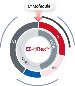
Fig 1. EZ-HRex™ System
III. Experimental Workflow for Constructing Point Mutation Cell Lines via the RNP Method
Step 1: gRNA Design
Design sgRNAs targeting the mutation site, giving preference to sequences where the cleavage site is within 10 bp of the mutation site. The design quality of the gRNA directly dictates subsequent cleavage efficiency and off-target risks. It is recommended to design 2–3 candidate gRNAs and pre-validate their cleavage efficiency.
Step 2: ssODN Template Synthesis
Synthesize single-stranded oligonucleotides (ssODNs) carrying the target mutation information to serve as HDR templates. These are typically 120–150 nt in length, with ends containing sequences homologous to the genome. Synonymous mutations can be introduced within the PAM site or gRNA region to prevent secondary cleavage.
Step 3: In Vitro Assembly of RNP Complex
Incubate chemically modified gRNAs with high-fidelity Cas9 protein (such as HiFi Cas9) in vitro to form stable RNP complexes. The assembly quality and molar ratio of the RNP complex directly impact editing efficiency.
Step 4: Co-Transfection (Electroporation/Lipofection)
Introduce the RNP complex and the ssODN template simultaneously into target cells via nuclear transfection technology (electroporation) or lipofection. Electroporation yields higher efficiency and is particularly suitable for hard-to-transfect cells. The advantages of the RNP method include non-reliance on DNA plasmids, transient expression, and low off-target risk.
Step 5: Monoclonal Screening
Perform monoclonal isolation post-transfection using methods such as counting dilution or limiting dilution. Given the complexity of gene editing events and the uncontrollable nature of heterozygous/homozygous states within mixed cell populations, monoclonal separation is mandatory for point mutation projects, regardless of whether the goal is a heterozygous or homozygous clone.
Step 6: Identification and Validation
Implement a three-tier screening system: "Monoclonal Screening + PCR Identification + Sequencing Validation." First, the target region is amplified via PCR to preliminarily identify positive clones. Subsequently, Sanger sequencing is performed for precise confirmation—this remains the gold standard for validating point mutations.
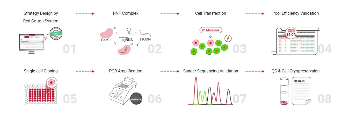
Fig 2. Workflow of Point Mutation Cell Line Construction by Ubigene
IV. Key Considerations for Constructing Point Mutation Cell Lines
(I) gRNA Design and Cleavage Site Selection
gRNA design serves as the opening move in point mutation construction and directly determines the success of the project. The closer the cleavage site is to the mutation site, the higher the HDR efficiency. If the mutation is too distant from the PAM site, consideration must be given to expanding the screening scope, changing the cleavage site, or adopting methods such as large-fragment recombination.
(II) Preventing Cas9 Secondary Cleavage
If an already edited site still retains the intact PAM sequence and gRNA recognition region, Cas9 may execute a secondary cleavage at that site. It is strongly recommended to introduce synonymous mutations within the PAM site or gRNA region in the ssODN template to disrupt the Cas9 recognition sequence, thereby significantly reducing the probability of secondary cleavage.
(III) Cell Status and Transfection Optimization
Low cell viability post-transfection is a common issue, with potential causes including excessive toxicity of transfection reagents, overly high transfection concentrations, or poor cell status. Solutions include: selecting appropriate transfection methods based on cell types (e.g., employing electroporation instead of lipofection for hard-to-transfect cells), optimizing the ratio of transfection reagents to plasmids, and ensuring the utilization of healthy cells in the logarithmic growth phase.
(IV) Pre-Assessment of Mutation Lethality
If a mutation occurs in essential genes, cell cycle-related genes, or mitochondrial function genes, it may impede cell growth, alter cell morphology, or even prevent the selection of homozygous mutant clones. A comprehensive assessment of the potential lethality of the mutation site should be conducted prior to the experiment; if necessary, generating heterozygous mutations is advisable.
(V) Single-Clone Screening Cannot Be Omitted
Single-clone isolation is mandatory for point mutation cell construction for three reasons:
- ① The heterozygous/homozygous status within mixed clones is uncontrollable, which compromises subsequent functional studies;
- ② Even within a single clone, significant variations can arise between different clones due to off-target effects;
- ③ Reliable positive clones can only be delivered after single-clone screening combined with sequencing validation.
(VI) Rational Combination of Validation Methods
Sanger sequencing stands as the gold standard for point mutation validation, yet relying solely on sequencing may overlook certain complex genotypes (such as compound heterozygous mutations). It is recommended to pair it with allele-specific PCR (ARMS-PCR) or qPCR for rapid preliminary screening, followed by sequencing for final confirmation. If necessary, NGS can be supplemented for off-target analysis.
(VII) Timeline Estimation and Planning
The construction cycle of point mutation cell lines is directly correlated with cell doubling time. Taking cells with a doubling time of under 24 hours as an example, conventional CRISPR editing methods typically require 10–12 weeks or even longer. Conversely, utilizing Ubigene’s EZ-HRex™ system, which substantially enhances HDR efficiency, allows for the delivery of validated positive mutant clones within 6–8 weeks , significantly shortening the project timeline.
Summary
The construction of point mutation cell lines represents an indispensable facet of functional genomics research. From dissecting the mechanisms of tumor driver mutations to precision modeling of hereditary diseases and screening target drug sensitivities, point mutation models play a pivotal role.
With advantages such as ease of operation, low off-target risk, and broad adaptability , the RNP method has become the mainstream technical route for point mutation cell line construction. However, persistent challenges like low HDR efficiency and heavy screening workloads have long hindered many researchers.
Building upon our original EZ-editor™ gene editing technology, Ubigene has innovatively introduced U⁺ molecules to develop the proprietary EZ-HRex™ technology . This technical upgrade effectively regulates the cell cycle and suppresses NHEJ activity, boosting HDR efficiency at the cell pool level to as high as 84% . Currently validated in various cell lines, including HEK293, iPSCs, HuH-7, and THP-1, our point mutation cell services offer a rapid delivery cycle of as fast as 6 weeks .
Ubigene Point Mutation Services
If you are planning an experiment for point mutation cell line construction, feel free to connect with us. Whether it's regarding protocol design, technical details, or timeline estimation, our professional team is here to provide insights and answers. Contact us to learn more >>>
 Promotions


