Commonly Used Cell Models and Their Applications in Melanoma Research


Commonly Used Cell Models and Applications in Melanoma Research
Melanoma, the "stealth killer" among skin cancers, has long been a focal point and a significant challenge in cancer research due to its high invasiveness and rapid metastatic capabilities. In this battle against melanoma, cell models have emerged as a powerful tool for scientists. They have not only helped to unveil the mysteries of melanoma but also serve as a crucial bridge from laboratory research to clinical therapies. Today, let's delve into the world of these models and explore how they are driving breakthroughs in melanoma research!
Contents
Human Malignant Melanoma Cell Line (A-375)
Ubigene Catalog #: YC-C036
Source: Derived from the skin tissue of a 54-year-old female patient with melanoma, this cell line is one of a series established by Giard DJ et al.
Characteristics:
- Epithelial-like morphology with adherent growth.
- Hypotriploid karyotype with 62 chromosomes.
- Tumorigenic in immunosuppressed mice and capable of forming colonies in agar.
- Widely used for studying the biological properties of melanoma, drug screening, and gene expression analysis.
Application Case: This cell line is used to study the metastatic mechanisms and drug responses of melanoma. When fluorescently labeled (e.g., with EGFP), it can be used for in vivo imaging and drug screening experiments. Tumor suppressor TP53 is considered one of the key regulators mediating the invasiveness and progression of melanoma. Over 80% of melanoma patients harbor wild-type (WT) but dysfunctional p53. A research team used the A375 (WT p53) human melanoma cell line to investigate the effects of JW-1-283. MTS and colony formation assays revealed that JW-1-283 inhibits the proliferation of melanoma cells and has a significant inhibitory effect on WT p53. Subcutaneous injection of A375 cells into the abdomen of NOD scid gamma (NSG) mice demonstrated that JW-1-283 significantly inhibits tumor growth. The research team is exploring the restoration of p53 function in melanoma patients as an alternative therapeutic strategy.
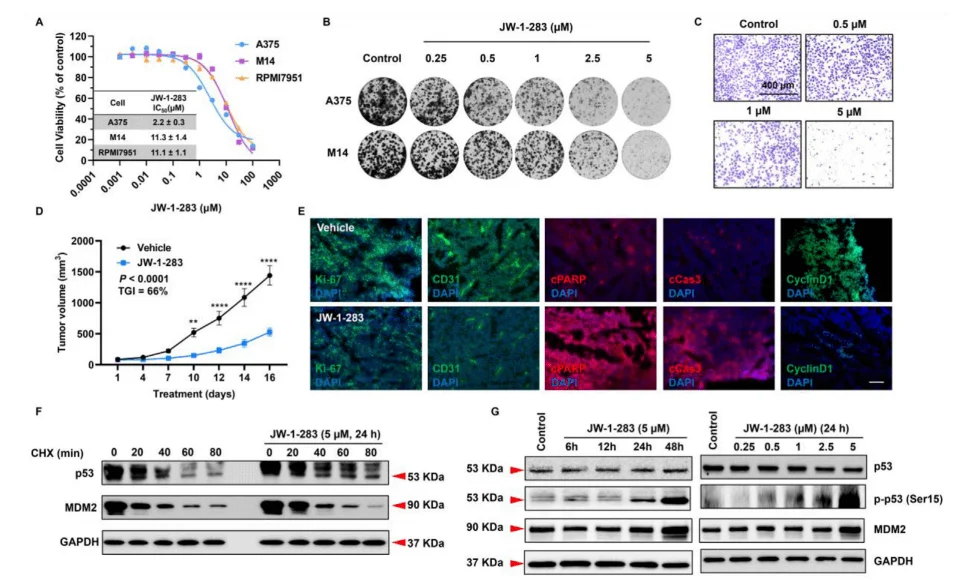
Figure 1. JW-1-283inhibits melanoma tumor growth by stabilizing the P53 pathway
Hot KO Cells-Melanoma Research
| PRAME Knockout cell line (A375) | YKO-H1073 |
| USP7 Knockout cell line (A-375) | YKO-H049 |
| B2M Knockout cell line (A-375) | YKO-H1281 |
| MAGEA4 Knockout cell line(A-375) | YKO-H274 |
| HLA-A Knockout cell line (A-375) | YKO-H1439 |
| Tgfb3 Knockout cell line (B16-F10) | YKO-M026 |
| Ythdf1 Knockout cell line (B16-F10) | YKO-M043 |
| F3 Knockout cell line (B16-F10) | YKO-M052 |
| Ttc14 Knockout cell line (B16-F10) | YKO-M053 |
| Klf12 Knockout cell line (B16-F10) | YKO-M057 |
Explore more KO cells, feel free to contact us!
Mouse Skin Melanoma Cell Line(B16-F10)
Ubigene Catalog #: YC-A011
Source: Derived from a highly metastatic subline of the mouse B16 melanoma cell line,originating from a spontaneous skin tumor in C57BL/6 mice.
Characteristics:
- Morphology is a mixture of epithelial-like and spindle-shaped cells with adherent growth.
- High metastatic potential,particularly to the lungs.
- Secretes large amounts of melanin,commonly used as a marker for cell proliferation and tumor growth.
Application Case: Utilized for studying the metastatic mechanisms of melanoma and immunotherapy. When labeled with luciferase, it can be used for in vivo imaging experiments and drug screening. This study investigated the impact of non-alcoholic steatohepatitis (NASH)-induced changes in the vascular niche on melanoma liver metastasis. B16F10 Luc2 cells were intrasplenically injected into Gata4 gene knockout mouse models, followed by splenectomy to study melanoma metastasis. The findings revealed that hepatic fibrosis promotes melanoma liver metastasis.
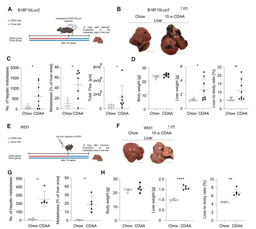
Figure 2. Steatohepatitis-Induced Changes in the Vascular Niche Promote Melanoma Metastasis
Human Melanoma Cell Line(SK-MEL-2)
Ubigene Catalog #: YC-A079
Source: Derived from the skin tissue of a 61-year-old male with malignant melanoma, established by the Memorial Sloan-Kettering Cancer Center(MSKCC).
Characteristics:
- Epithelial-like morphology with adherent growth.
- Complex karyotype with high genetic heterogeneity typical of melanoma.
- High proliferation and invasive potential,with resistance to certain chemotherapeutic and targeted therapies.
Application Case: Commonly used to study signaling pathways (e.g.,MAPK/ERK) and the impact of gene mutations (e.g.,BRAF,NRAS) on tumor growth and metastasis. A research team conducted xenograft studies with SK-MEL-2 cells (NRASQ61R) and found that STK19 (serine/threonine kinase) promotes NRAS-driven melanoma malignancy by phosphorylating serine 89 (S89) of NRAS. They also discovered that a small-molecule inhibitor of STK19 (ZT-12-037-01(1a)) effectively inhibits NRAS-driven melanoma progression, highlighting its potential as a targeted therapy for NRAS-driven melanoma.
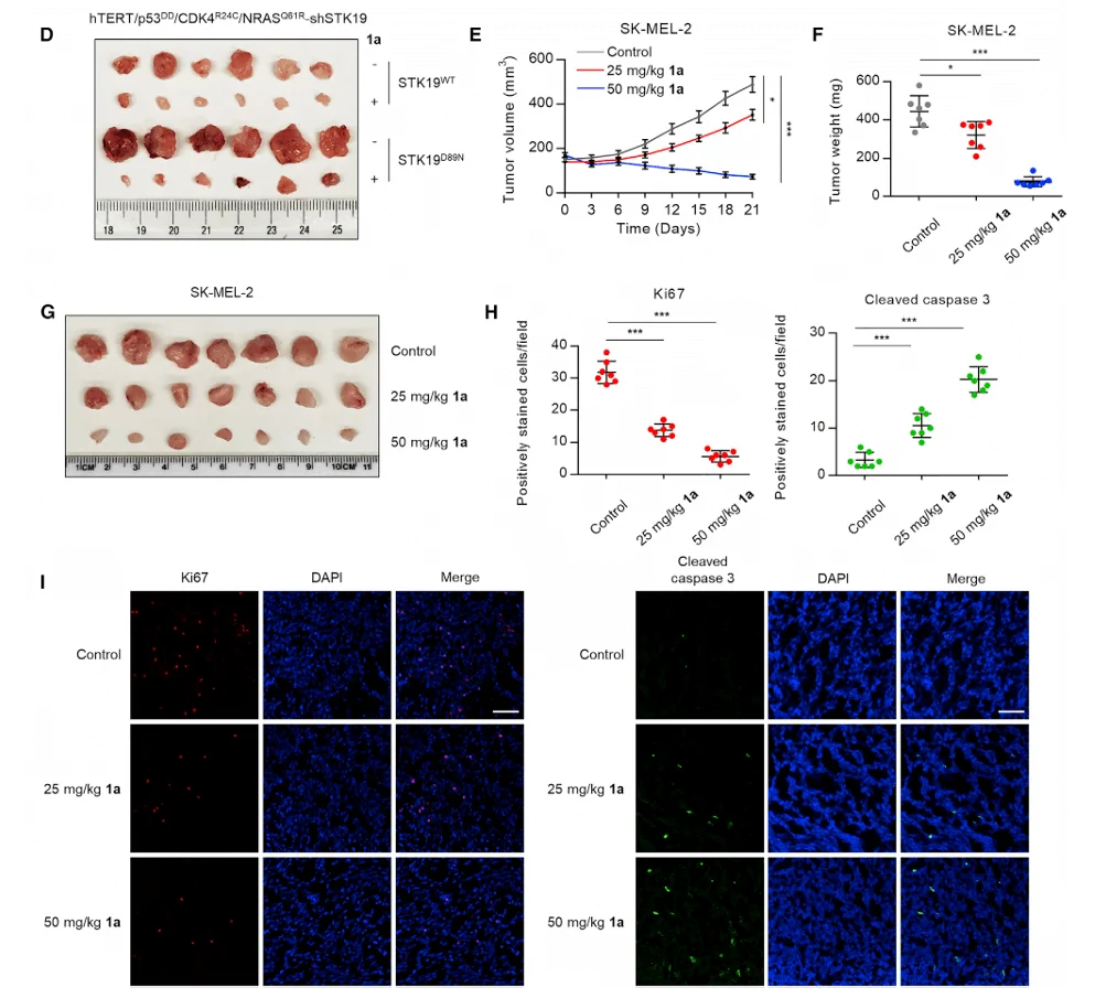
Figure 3. ZT-012-037-1(1a) inhibits the development and growth of melanoma driven by oncogenic NRAS.
Summary
Every step in melanoma research relies on the support of these models. From basic cell line models to gene-edited models, scientists continue to explore new methods to better understand and treat melanoma. These models not only help us gain a deeper understanding of the biological behavior of melanoma but also serve as a crucial experimental basis for developing new therapeutic approaches.
Ubigene offers gene editing services (KO/KI/PM) and stable cell line customization for melanoma research-related cells. Inquiries are welcome!
Reference:
[1] Xie Y, Hou R, Hartman KL, Jiang J, Wu Z, Li W. JW-1-283 inhibits melanoma tumor growth via stabilization of the p53 pathway. Genes Dis. 2023 Jul 17;11(3):101036. doi: 10.1016/j.gendis.2023.06.009. PMID: 38274379; PMCID: PMC10808915.
[2] Hoffmann, J., Schüler, J., Dietsch, B. et al. Steatohepatitis-induced vascular niche alterations promote melanoma metastasis. Cancer Metab 13, 5 (2025). https://doi.org/10.1186/s40170-025-00374-6
[3] Yin C, Zhu B, Zhang T, Liu T, Chen S, Liu Y, Li X, Miao X, Li S, Mi X, Zhang J, Li L, Wei G, Xu ZX, Gao X, Huang C, Wei Z, Goding CR, Wang P, Deng X, Cui R. Pharmacological Targeting of STK19 Inhibits Oncogenic NRAS-Driven Melanomagenesis. Cell. 2019 Feb 21;176(5):1113-1127.e16. doi: 10.1016/j.cell.2019.01.002. Epub 2019 Jan 31. PMID: 30712867.
 Promotions


