UBIGENE

Comprehensive CRISPR Gene Editing Solutions to Power Your Research: KO Cell Lines, CRISPR Screen & More

CRISPR Gene Knockin Cell Line Service

Start at $5980 | 10 Wks deliver homozygous clone | HDR up to 84%

Chat with an Expert

Gene Knockin Cell Line

Cells are co-transfected via nucleofection with a gRNA expression vector (carry Cas9) and a donor template carrying the insert of interest. A site-specific DNA double-strand break (DSB) is introduced at the target locus, and precise knock-in is achieved through homology-directed repair (HDR) using the donor as a template. Following antibiotic selection, single-cell clones are isolated (fluorescence can be used for preliminary screening if applicable). Targeted PCR and Sanger sequencing are performed to confirm correct integration, and validated positive clones are delivered with a complete data report.

KI Cell Line Service Highlights
High gene cleavage and homologous recombination efficiency, achieving a 10-20x increase in editing efficiency; HDR efficiency up to 84%.
Extensive track record, with over 10,000 successful gene editing projects across more than 300 cell types.
Rapid turnaround: monoclonal knock-in cell lines delivered in as fast as 10 weeks.

Gene Knockin Cell Line Service Detail

Service Type

Gene Knockin

Cell Line Type

Tumor Cells, Immortalized Cell Lines. Various cell types, including iPS cells

Project Price (USD)

From $5980

Turnaround

As fast as 10 weeks

Deliverables

1. ≥1 homozygous Single Clone(>106 cells/vial, 2 vials per clone) 2. Wild-Type Cells(>106 cells/vial, 2 vials) 3. Experimental Reports

Knockin Validation

PCR(Standard), Sanger sequencing(Standard)

* For research use only. Not intended for human or animal clinical trials, therapeutic or diagnostic use.
300+ Successful Gene-editing Cell Line Types

Digestive System

Human Gallbladder Carcinoma Cell Line (GBC-SD)Human Hepatoma Cell Line (Hep G2)Human Hepatoma Cell Line (SNU-387)Human Hepatoma Cell Line (HuH-7)Human Hepatoma Cell Line (SK-HEP-1)Human Hepatoma Cell Line (Hep 3B)Human Normal Hepatocytes Cell line (L-02)Human Colon Cancer Cell Line (HCT 116)Human Colon Cancer Cell Line (HT-29)Human colorectal adenocarcinoma Cell Line (RKO)Human colorectal adenocarcinoma Cell Line (Caco-2)Human Esophageal Squamous Carcinoma Cell Line (KYSE-150)Human gastric cancer Cell Line (AGS)Mouse Hepatocarcinoma Cell Line (Hepa 1-6)Murine colorectal carcinoma Cell Line (CT26.WT)Murine colorectal carcinoma Cell Line (MC38)Human Esophageal Squamous Carcinoma Cell Line (KYSE-30)Mouse Hepatocarcinoma Cell Line (H22)Human Gastric Cancer Cell Line (NCI-N87)Human colorectal adenocarcinoma Cell Line (SW480)Human Hepatoma Cell Line (SMMC-7721)Porcine intestinal epithelial Cell Line (IPEC-J2)Human Gastric Cancer Cell Line (HGC-27)Human Gastric Cancer Cell Line (MGC-803)Human renal carcinoma cell line (ACHN)Human colorectal adenocarcinoma Cell Line (LS 174T)Immortalized Human Hepatic Cell Line (C3A)Human Hepatoma Cell Line (PLC/PRF/5)Human colorectal adenocarcinoma Cell Line (SW 48)Human colorectal adenocarcinoma Cell Line (SW948)Human colorectal adenocarcinoma Cell Line (NCI-H716)Mouse hepatocyte Cell Line (AML12)Human Gastric Cancer Cell Line (SGC-7901)Human Ileocecal Cancer Cell Line (HRT-18)Human Colon Cancer Cell Line (LoVo)Human Hepatoma Cell Line (MHCC97-H)Human Gastric Cancer Cell Line (MKN-45)Human Colon Cancer Cell Line (COLO 205)Rat Liver Cell Line (BRL)Mouse Gastric Cancer Cell Line (MFC)Human Colon Cancer Cell Line (SW620)Human Hepatoma Cell Line (Li-7)Mouse Ascites Sarcoma Cell Line (S180)Rat Liver Cell Line (BRL-3A)Human Colon Cancer Cell Line (HCT-15)Human Gastric Cancer Cell Line (KATO III)Human colorectal adenocarcinoma Cell Line (SW1463)Human Gastric Cancer Cell Line (SNU-5)Human colorectal adenocarcinoma Cell Line (SW1116 [SW 1116, SW-1116])Human Hepatic Stellate Cell Line (LX-2)Human Hepatoma Cell Line (HCCLM3)

Endocrine System

Human Submandibular Gland Epidermoid Carcinoma Cell Line (A-253)Human Mammary Epithelial Cell Line (MCF 10A)Human Breast Cancer Cell Line (MDA-MB-231)Human Breast Cancer Cell Line (MCF7)Human Breast Cancer Cell Line (MDA-MB-415)Human Breast Cancer Cell Line (MDA-MB-468)Human Breast Epithelial Cell Line Integrating SV40 Gene (HBL-100)Human Prostate Cancer Cell Line (LNCaP Clone FGC)Human Prostate Cancer Cell Line (PC-3)Human Prostate Cancer Cell Line (22RV1)Human pancreatic cancer Cell Line (BxPC-3)Mouse Breast Cancer Cell Line (4T1)Mouse Acinar Pancreatic Cell Line (266-6)Human Breast Cancer Cell Line (JIMT-1)Mouse prostate cancer Cell Line (RM-1)Human Breast Cancer Cell Line (SK-BR-3)Human Breast Cancer Cell Line (HCC1806)Human Thyroid Normal Cell Line (Nthy-ori 3-1)Mouse pancreatic cancer Cell Line (Pan-02)Human Pancreatic Carcinoma Cell Line (SW1990)Human acute lymphoblastic leukemia Cell Line (MOLT4)Human Pancreatic Carcinoma Cell Line (Panc-1)Mouse Breast Adenocarcinoma Cell Line (E0771)Human Thyroid Squamous Cell Carcinoma Cell line (SW579)Human Breast Cancer Cell Line (HCC1143)Human breast cancer Cell Line (BT-549)Human acute lymphoblastic leukemia Cell Line (MT-4)Py230 Cell Line (Py230)Human Thyroid Cancer Cell Line (TPC-1)Human Prostate Cancer Cell Line (PC-3M)Human Burkitt's Lymphoma Cell Line(RAJI)Human Metastatic Pancreatic Adenocarcinoma Cell Line (AsPC-1)Human NK Leukemia Cell Line (YT)Human Prostate Cancer Cell Line (DU145)Mouse Mammary Epithelial Cell Line (HC11)Mouse Breast Cancer Cell Line (C127)Human Thyroid Normal Cell Line (Nthy-ori 3-1)Human Pancreatic Carcinoma Cell Line (MIA PaCa-2)Mouse Breast Cancer Cell Line (EMT6)Human Prostate Cancer Cell Line (VCaP)Human Breast Cancer Cell Line (SUM-149PT)Human breast ductal cancer cell Line (BT474)Human Pancreatic Carcinoma Cell Line (Capan2)

Respiratory System

Human Lung Cancer Cell Line (A549)Human Lung Cancer Cell Line (NCI-H1299)Human Lung Cancer Cell Line (NCI-H520)Human Lung Cancer Cell Line (SK-MES-1)Mouse Lewis lung carcinoma Cell Line (LLC)Human Bronchial Epithelial Cell Line (BEAS-2B)Human Lung Cancer Cell Line (HCC827)Porcine alveolar macrophage cell line (3D4/21)Human lung adenocarcinoma Cell Line (PC9)Human Lung Cancer Cell Line (NCI-H1650)Human Lung Cancer Cell Line (NCI-H727)Human lung adenocarcinoma Cell Line (NCI-H1975)Human lung cancer Cell Line (NCI-H292)Human Lung Cancer Cell Line (NCI-H23)Human Lung Cancer Cell Line (MSTO-211H)Human Lung Cancer Cell Line (NCI-H358)Human Non-small Cell Lung Carcinoma Cell Line (NCI-H460)Human lung adenocarcinoma Cell Line (Calu-3)Human Fetal Lung Fibroblast Cell Line (MRC-5)Human Highly Metastatic Lung Cancer Cell Line (95-D)Human Colon Carcinoma Cell Line (T84)MLE-12 Cell Complete Medium (MLE-12)Human Fetal Lung Fibroblast Cell Line (WI-38)Human Lung Cancer Cell Line (DMS114))Human lung adenocarcinoma Cell Line (NCI-H1373)Human Lung Cancer Cell Line (NCI-H1703)Human Lung Cancer Cell Line (DMS 53)Human Small Cell Lung Cancer Cell Line (NCI-H82 [H82])Human Small Cell Lung Cancer Cell Line (SHP-77)Human Lung Cancer Cell Line (NCI-H2170)

Reproductive System

Human Amniotic Cell Line (WISH)Human Highly Metastatic Ovarian Cancer Cell Line (HO-8910PM)African green monkey kidney Cell Line (Marc-145)Human Cervical Carcinoma Cell Line (Hela)Human ovarian cancer Cell Line (OVCAR-3)Human Ovarian Adenocarcinoma Cell Line (Caov-3)Mouse Embryonic Fibroblasts (NIH/3T3)Mouse Fibroblast Cell Line (TM3)Human Villous Trophoblast (HTR-8/SVneo)Human Ovarian Granulosa Cell Line (KGN)Chinese Hamster Ovary Cell Line (CHO-K1)Human Endometrial Adenocarcinoma Cell Line (MFE-280)Mouse ovarian epithelial cancer Cell (ID8)Human endometrial adenocarcinoma Cell (HEC-1-B)Human Endometrial Carcinoma Cell Line (ISHIKAWA)Human Cervical Carcinoma Cell Line (C-33 A)Human Cervical Carcinoma Cell Line (Ca Ski)Human Endometrial Carcinoma Cell Line (KLE)Mouse Teratocarcinoma Cell Line (P19)Human Ovarian Clear Cell Carcinoma Cell Line (ES-2)Human Endometrial Adenocarcinoma Cell Line (AN3 CA)Human Cervical Carcinoma Cell Line (hela 229)Human Ovarian Adenocarcinoma Cell Line (SK-OV-3)Human Choriocarcinoma Cell Line (Bewo)

Circulatory System

Rat Cardiac Myocytes (H9c2(2-1))Mouse Myoblast Cell Line (C2C12)Human Cardiac Myocytes (AC16)Mouse Aortic Smooth Muscle Cell Line (MOVAS)

Blood and lymphatic System

Mouse Hybridoma Cell Line (AE-1)Hybridoma (Anti-Cd3) Cell Line (OKT 3)Human Monocytic Cell Line (THP-1)Human Histiocytic Lymphoma Cell Line (U-937)Human T Lymphocyte Cell Line (Jurkat, Clone E6-1)Human Acute Lymphocyte Cell Line (OCI-Aml-3)Human myelogenous Leukemia Cell Line (K-562)Mouse Macrophage Cell Line (RAW 264.7)Human Acute Promyelocytic Leukemia Cell Line (HL-60)Human B Lymphoma Cell Line (U-2932)Mouse B Lymphoma Cell Line (A20)Human Myelomonocytic Leukemia Cell Line (MV4-11)Human B Lymphoma Cell Line (SU-DHL-4)Human Acute Myeloid Leukemia Cell Line (KG-1a)Human Erythroleukemia Cell Line (HEL)Mouse Pro B Cell Line (BAF3)Human acute promyelocytic leukemia cell line (NB4)Human T-lymphocyte Cell Line (Hut-78)Human B Lymphocytes Cell Line (GM12878)Mouse Lymphoma Cell Line (EL4)Mouse Macrophage Cell Line (Ana-1)Human Erythroleukemia Cell Line (TF-1)Human Lymphoma Cell Line (BC-3)Human Multiple Myeloma Peripheral Blood B Lymphocytes (RPMI 8226)Human Burkitt's Lymphoma B Lymphocytes (Ramos)Human B lymphoma cell line (SU-DHL-6)Human Acute Lymphoblastic Leukemia Cell Line (Non-B Non-T)(Reh)Human Acute Lymphocyte Cell Line (Kasumi-1)Human Mantle Cell Lymphoma Cell Line (JeKo-1)Human peripheral blood mantle lymphoma Cell Line (Mino)Human Acute Myeloid Leukemia Cell Line (MOLM13)Human B lymphocytic leukaemia Cell Line (NALM-6)Human B lymphoma Cell Line (SU-DHL-8)

Brain and Nervous System

Rat Glioblastoma Cell Line (C6)Mouse Hippocampal Neuronal Cell Line (HT-22)Human Neuroblastoma Cell Line (SK-N-SH)Human Neuroblastoma Cell Line (SH-SY5Y)Human glioblastoma Cell Line (U-87 MG)Human glioblastoma Cell Line (U251MG)Mouse Anterior Parietal Bone Cell Line (MC3T3-E1 Subclone 14)Mouse microglia Cell Line (BV2)Human microglial clone 3 Cell Line (HMC3)Mouse neuroblastoma Cell Line (Neuro-2a)Human glioblastoma Cell Line (LN-229)Mouse Glioblastoma Cell Line (GL261)Human Glioblastoma Cell Line (T98G)Human Glioblastoma Cell Line (A-172)Human Neuroblastoma Cell Line (SK-N-BE(2))Human Brain Astrocytoma Cell Line (Hs 683)Rat Glioblastoma Cell Line (RG2)

Urinary System

African green monkey kidney Cell (Vero)African green monkey kidney Cell (VERO C1008 (E6))Dog Kidney Cell Line (MDCK(NBL-2))Human Embryonic Kidney Cell Line (293T)Human Embryonic Kidney Cell Line (293T)Human Embryonic Kidney Cell Line (HEK293)Rat adrenal pheochromocytoma Cell Line (Low differentiation)(PC-12)Human Bladder Transitional Cell Carcinoma Cell Line (T24)Human bladder carcinoma cell line (5637)Mouse Bladder Cancer Cell (MB-49)Human renal cell carcinoma Cell Line (786-0)Human renal cell carcinoma Cell Line(MS751)Mouse podocyte Cell Line(MPC-5)Pig Kidney Cell line(PK-15)Human Embryonic Kidney Cell Line (2V6.11)Human Bladder Transitional Cell Carcinoma Cell Line (SW780)Transformed (SV40) African Green Monkey Kidney Cell Line (COS-1)Transformed (SV40) African Green Monkey Kidney Cell Line (COS-7)Rat Kidney Cell Line (NRK-52E)Mouse Renal Adenocarcinoma Cell Line (RenCa)Bovine Kidney (MDBK) Cell Line (MDBK (NBL-1))Hamster Kidney Fibroblasts (BHK-21)Human Renal Proximal Tubular Epithelial Cell Line (HK-2)Human Urinary Bladder Squamous Cell Carcinoma Cell Line (SCaBER)Mouse Renal Adenocarcinoma Cell Line (RAG)Mouse Bladder Cancer Cell (MBT-2)Human renal carcinoma Cell Line (OS-RC-2)

Skeleton, Articulus, Soft Tissue, Derma System

Human Umbilical Vein Cell Line (EA.hy926)Mouse Lewis lung carcinoma Cell Line (U-2 OS)Human Osteosarcoma Cell Line (Saos-2)Human skin squamous carcinoma Cell Line (A-431)Murine melanoma Cell Line (B16-F10)Human malignant melanoma Cell Line (A-375)Human Skin Fibroblast Cell Line (BJ)Mouse Fibroblast Cell Line (L-929)Mouse Lewis lung carcinoma Cell Line (SW 1353)Human fibrosarcoma Cell Line (HT-1080)Rat osteosarcoma Cell Line(UMR-106)Human Immortalized Keratinocytes Cell Line (HaCaT)Chicken Fibroblast Cell Line (DF1)Human Multiple Myeloma Cell Line (MM.1S)Human Corneal Epithelial Cell Line (HCE-T)(HCE-T)Immortalized Human Lens Epithelial Adherent Cell Line (SRA01/04)Mouse Lewis lung carcinoma Cell Line (MG-63)Mouse Osteosarcoma Cell Line (K7M2-WT)Mouse Lewis lung carcinoma Cell Line (143B)Mouse Subcutaneous Connective Tissue Cell Line (A9)Mouse squamous carcinoma Cell Line (SCC-7)Human Choriocarcinoma Cell Line (JAR)Human bone osteosarcoma epithelial Cell Line (KHOS-240S)Mouse Chondrogenic Cell Line (ATDC5)

Stem Cell Lines

Human induced pluripotent stem cells (iPSC) (IPSC-DYR0100)

Ocular, Otolaryngologic and Oral System

Human Pharyngeal Carcinoma Cell Line (Pleural Effusion Metastasis) (Detroit 562)Human retinal pigment epithelial Cell Line (ARPE-19)Human Nasopharyngeal Carcinoma Cell Line (CNE1)Human oral squamous carcinoma Cell Line (HSC3)Human nasopharyngeal carcinoma Cell Line (HK-1)Human oral squamous carcinoma cell line (CAL-27)Mouse Cochlear Hair Cell Line (HEI-OC1)Human Retinoblastoma Cell Line (Y79)Human Tongue Squamous Carcinoma Cell Line (SCC-9)Human Pharyngeal Squamous Cell Carcinoma Cell Line (FaDu)

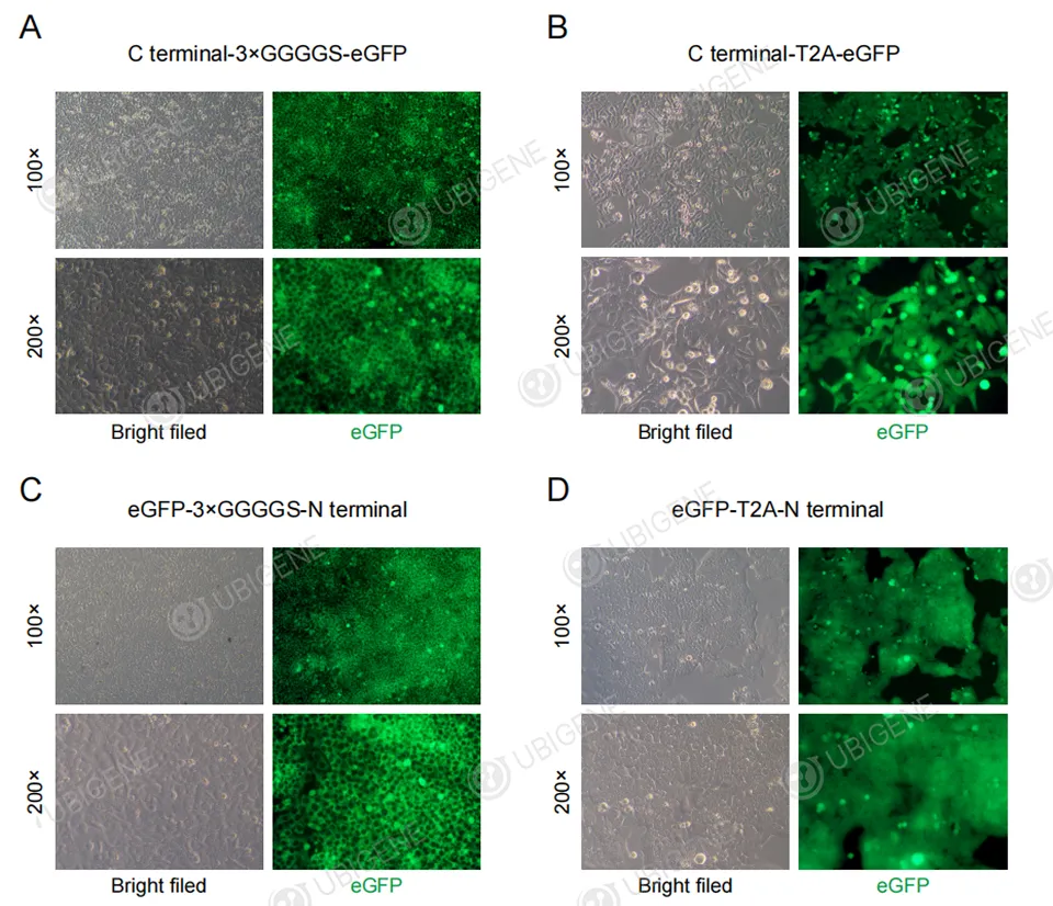
Gene Knockin (KI) Cells Construction Methods

Protein fusion

View Picture

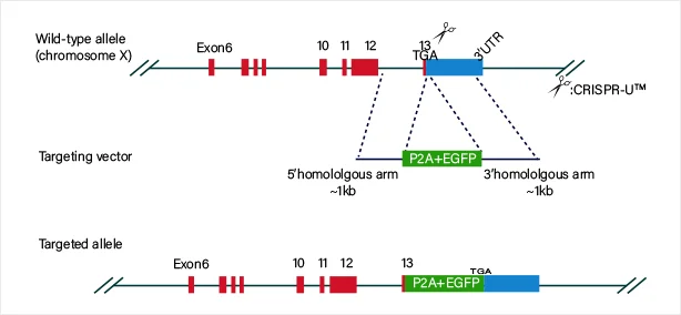
The guide RNA and Cas9 complex induce a double-strand break (DSB) at the target site of the DNA. The donor vector carrying the knock-in sequence serves as the template for homologous recombination repair (HDR), facilitating the recombination of the knock-in sequence into the target site.

Replacement of specific locus

View Picture

Workflow and Validation

01

Strategy Design by Red Cotton System

02

RNP Complex

03

Cell Transfection

04

Pool Efficiency Validation

05

Single-cell Cloning

06

PCR Amplification

07

Sanger Sequencing Validation

08

QC & Cell Cryopreservaion

Case Study

Figure 1: EEF1A1 knockin A549 cell pool was generated using the EZ-HRex™ technique, with an HDR efficiency reaching 88%.
(A)
(B)
Figure 2. CRISPR gene knock-in in A549 cells. Cas9/gRNA and oligo were delivered into A549 cells via electroporation to achieve targeted gene knock-in. (A) Sanger sequencing validation of the knock-in at the target locus.(B) Genotyping PCR analysis of monoclonal cell lines with homozygous knock-in.

Research Review

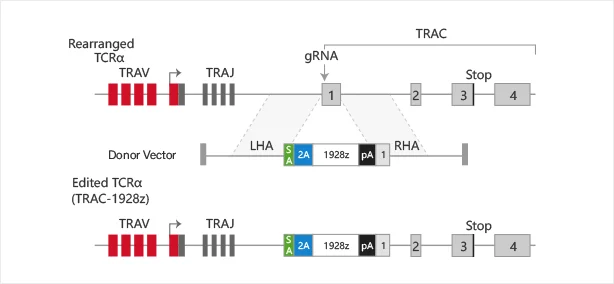
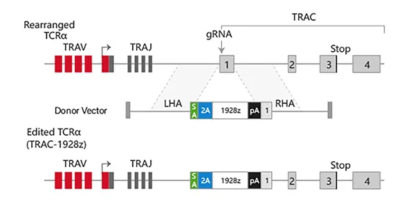
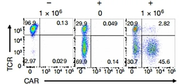
Targeting a CAR to the TRAC locus with CRISPR/Cas9 enhances tumour rejection

T cells expressing chimeric antigen receptors (CARs) can mediate tumor rejection and are an effective therapy for B-cell malignancies. By applying site-specific genome editing, a CD19-specific CAR cassette can be precisely integrated into the TRAC locus in T cells via donor template–mediated homologous recombination. This strategy enables uniform CAR expression in primary human peripheral blood T cells and enhances their immune response capacity.

View Picture

Set up multiple infection gradients with different MOIs to test viral infection efficiency. Calculate the corresponding cell infection rates and select the MOI that achieves approximately 30% infection efficiency as the condition for library virus infection.

View Picture

TCR/CAR Flow Cytometry Analysis When Cas9, gRNA, and donor template are co-delivered, CAR expression can be detected as early as 4 days post-editing by flow cytometry, indicating successful knock-in at the target locus.

FAQs

1. What is CRISPR knock-in?

CRISPR knock-in refers to the process of using the CRISPR/Cas9 gene-editing technology to insert a specific genetic sequence (such as a gene, regulatory element, or mutation) into a precise location in the genome. This technique is used to add functional genes or introduce specific genetic modifications in a targeted manner, as opposed to CRISPR knock-out, where genes are disrupted or deleted.

2. How does CRISPR Knock-in Work?

  • Designing the Guide RNA (gRNA): A guide RNA is designed to specifically target a location in the genome where the new DNA sequence will be inserted. This RNA sequence is complementary to the target DNA sequence, ensuring that Cas9 cuts the correct location in the genome.
  • Cas9 Protein: The Cas9 protein, which is part of the CRISPR system, is responsible for making a double-strand break at the target location in the DNA.
  • Donor DNA Template: To achieve the knock-in, a donor DNA template is introduced into the cell. This template contains the genetic sequence that will be inserted into the genome. The donor template typically has homology arms (sequences that match the regions adjacent to the target break) to guide the cell's repair machinery.
  • Homology-Directed Repair (HDR): After Cas9 cuts the DNA, the cell uses its HDR mechanism to repair the break. If a donor template is provided, the cell incorporates the new genetic material from the donor DNA into the genome at the break site, which results in the insertion of the desired sequence.

3. What are the differences between CRISPR gene knock-in and knock-out ?

key Differences
FeatureCRISPR Knock-inCRISPR Knock-out
Purpose To insert a specific gene or sequence into the genome. To disrupt or inactivate a gene in the genome.
Outcome Addition of a gene or sequence at a specific site. Loss-of-function mutation due to gene disruption.
Repair Mechanism Homology-directed repair (HDR) using a donor template. Non-homologous end joining (NHEJ) that introduces errors at the break site.
Resulting Change Insertion of new genetic material (e.g., a gene, mutation, or reporter sequence). Gene is disrupted or inactivated, often with indels.
Use Cases Gene therapy, disease modeling, synthetic biology, protein production. Gene function studies, disease modeling, cancer research, drug discovery.

4. What are the applications of CRISPR Knock-in?

  • Gene Therapy: Knock-in CRISPR can be used to insert healthy copies of defective genes in patients with genetic disorders. For example, correcting mutations in the CFTR gene (responsible for cystic fibrosis) by inserting a functional copy into a patient's cells.
  • Disease Modeling: Researchers use knock-in to introduce specific mutations into the genome of animal models (like mice or rats) to study diseases or test therapies. This helps in creating models of genetic diseases such as cancer or neurodegenerative conditions.
  • Synthetic Biology: Scientists can insert new genes into organisms (bacteria, yeast, or plants) to create new functions, such as engineered microbes that produce biofuels or therapeutic proteins.
  • Drug Development: In drug discovery, knock-in CRISPR is used to create cell lines or animals that express specific mutations or diseases, allowing for more accurate testing of drugs or potential treatments.
  • Cell Line Engineering: For industrial or therapeutic use, CRISPR knock-in can generate cell lines that express desired proteins, enzymes, or other bioactive molecules for research or commercial purposes.

5. How to perform a CRISPR Knockin Experiment?

  • Cell Type: Choose an appropriate cell line or primary cell type for your experiment.
  • Efficiency: CRISPR knock-ins generally have lower efficiency than knock-outs, and larger inserts can reduce efficiency further.
  • Delivery Method: Choose a delivery method (electroporation, lipid-based, viral) based on your cell type's transfection efficiency.
  • Homology-directed Repair (HDR): The success of knock-ins heavily relies on HDR, which is not as efficient as non-homologous end joining (NHEJ). You can enhance HDR by using HDR enhancers or small molecules (like SCR7).
  • Verification: Always validate your knock-in using multiple methods (PCR, sequencing, protein expression).

Ubigene could provide 4 solutions to meet different mutation needs! Get in touch with our experts now

6. How to validate CRISPR knockin cell?

Validating CRISPR knock-in cells involves confirming:

  • Correct insertion of the desired gene at the target location (via PCR, sequencing, Southern blot).
  • Expression of the inserted gene at the RNA and protein levels (via RT-PCR, Western blot, fluorescence assays).
  • Functionality of the inserted gene, especially for functional knock-ins (via fluorescence or activity assays).
  • Absence of off-target effects, to ensure the edit is precise (via sequencing or specific assays).

7. What are the challenges and limitations of CRISPR Knock-in?

After years of R&D of Ubigene, based on the original EZ-editor™ gene-editing technique, Ubigene has upgraded it to EZ-HRex™ New Technique. With the new technology, the proportion of HDR genotypes can reach up to 84%. Get in touch with our experts now

  • Efficiency: The efficiency of knock-in can be low, especially compared to knock-out techniques, because homology-directed repair (HDR) is less common than the alternative repair pathway, non-homologous end joining (NHEJ), which often leads to deletions or errors rather than precise insertions.
  • Off-target Effects: Although CRISPR technology is highly specific, there is still the potential for off-target edits, where the CRISPR machinery cuts DNA at unintended sites, leading to unwanted genetic changes.
  • Delivery: Delivering the CRISPR/Cas9 components (guide RNA, Cas9, and donor DNA) into cells, especially in vivo (in living organisms), is challenging. Efficient delivery systems are crucial for the success of CRISPR knock-in applications.

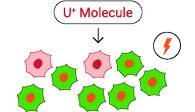
8. How to increase knock-In efficiency?

Ubigene has revolutionized gene editing with the new EZ-HRex™ technique, now enhanced with the innovative U+ Molecule. This breakthrough allows HDR genotype proportions to reach up to 84% at the post-transfection cell pool level. Additionally, Ubigene offers 4 tailored solutions to address diverse mutation requirements, ensuring maximum flexibility and precision in your experiments! Contact us now

9. How much does CRISPR Knock-in cost?

Ubigene offers global gene knock-in services, starting at just $6,480!

Contact Us

Simply fill out the form below to leave your inquiry
— we will respond within 24 Hours

* Name

* Email

* Institution

* Products & Services of Interest

If email is not available, how else can we reach you?

How did you hear about us?

Logo
E-mail: info@ubigene.com
WhatsAPP: +86 153 6067 3248
TEL: +86 153 6067 3248(Int'l)
Copyright © 2025 Ubigene. All rights reserved.