Luciferase Assay: Principles, Purpose, and Process


I. Technical Principle of Luciferase Reporter Gene Assays
Luciferases are a class of enzymes capable of catalyzing the oxidation of luciferin to generate bioluminescent signals. Common luciferases include Firefly luciferase (FLuc), Renilla luciferase (RLuc), and Gaussia luciferase (GLuc). The luciferase reporter gene assay technology is widely utilized in studies concerning gene expression regulation and high-throughput drug screening.
Taking the luciferase reporter assay system as an example: the promoter or regulatory sequence of a target gene is cloned upstream of the luciferase gene. Following cell transfection, the addition of a substrate (such as D-luciferin) generates a luminescent signal, where the luminescence intensity is directly proportional to the transcriptional activity of the gene.
Unlike Green Fluorescent Protein (GFP), luciferase assay detection does not require external excitation light and is not endogenously expressed in most cells, resulting in an extremely low background. Furthermore, it features strong light penetration, a wide dynamic range, and high sensitivity. Due to its ultra-high sensitivity and wide dynamic range, luciferase-based reporter assays are extensively used, making the luciferase reporter system an ideal quantitative detection tool.
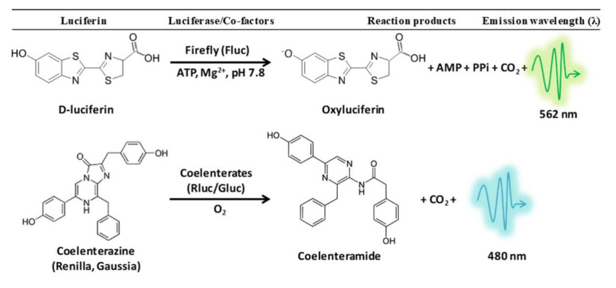
Figure 1: Schematic diagram of the luciferase reporter gene assay principle
Single vs. Dual Reporter Gene Assay Systems
- Single Reporter Gene Assay System: This system detects the activity of only one luciferase. While its operating steps are relatively simple, it is highly susceptible to interference from sample variations such as cell number or protein content. Consequently, data normalization based on cell count or protein concentration is typically required during experiments.
- Dual-Luciferase Reporter Gene Assay System: This dual-luciferase reporter assay system involves the co-transfection of two luciferase plasmids (such as FLuc and RLuc). Firefly luciferase (FLuc) is used to detect the experimental signal, while Renilla luciferase (RLuc) serves as an internal control. The two enzymes utilize different substrates to produce light signals at distinct wavelengths, effectively correcting for variations in transfection efficiency and cell viability. Combining the advantages of both luciferases, this system is widely adopted for precise detection.
II.Comparison of Common Reporter Gene Systems
Common reporter genes include β-galactosidase (LacZ), chloramphenicol acetyltransferase (CAT), fluorescent proteins (such as GFP), and luciferases, each suited for specific scenarios.
- CAT assays offer high sensitivity but involve tedious operations.
- LacZ is commonly used as an internal control for transfection.
- GFP allows for the real-time observation of living cells, but it requires external excitation light and faces significant interference from tissue autofluorescence.
In contrast, the luciferase reporter gene relies on a highly specific substrate-enzyme reaction to emit light, requiring no excitation light and yielding low background noise. Additionally, the luciferase signal is stable with a wide dynamic range and rapid response time, making it easy to adapt for high-throughput screening (HTS) and multiplexed reporter assays. Therefore, it is widely applied in gene expression regulation, cell tracking, and drug screening.
| Reporter Gene | Detection Method / Substrate | Advantages | Typical Applications |
| Luciferase | Luminescence emitted after luciferin excitation | High sensitivity, rapid response, no excitation light needed, low background | Transcriptional regulation studies, in vivo cell tracking, drug screening |
| Fluorescent Protein (GFP, etc.) | Fluorescence emitted via 488 nm excitation light | Enables real-time observation of cell/protein distribution | Cell tracking, localization (long-term observation) |
| β-galactosidase (LacZ) | Colorimetric readout via the decomposition of O-nitrophenyl-β-D-galactopyranoside | Easy to detect | Transfection efficiency correction, internal control |
| CAT | Acetyl-CoA reacts with chloramphenicol to produce acetylated chloramphenicol | Sensitive, low background | Gene expression determination (outdated method) |
Table 1: Comparative Analysis of Different Reporter Gene Systems
III. Construction and Application of Luciferase-Labeled Stable Cell Lines (Luc Stable Cell Line Services)
-
Experimental Purpose
Constructing luciferase-labeled stable cell lines (Luc stable cell lines) enables the real-time monitoring of gene expression or cellular behavior. They are frequently used to study transcription factor-promoter interactions, miRNA-target gene interactions, signaling pathway activation, and the screening of drugs or small molecules. -
Experimental Steps
-
Plasmid Construction
When studying the effects of transcription factors on gene expression, the pGL4.20 vector and pRL vector series are commonly chosen. The pGL4.20 vector lacks a promoter upstream of the FLuc gene, providing a multiple cloning site (MCS) where the promoter sequence of interest can be inserted. The pRL series vectors differ primarily by their promoters; an appropriate promoter can be selected based on experimental needs, with the weaker TK promoter being the standard choice.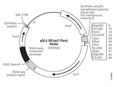
Figure 2: Schematic diagrams of the pGL4.20 vector and pRL vector series
When studying microRNA and mRNA interactions, pMIR-REPORT Luciferase can be used. This vector features an MCS at the 3' UTR of FLuc, allowing the insertion of the target gene's 3' UTR. The internal control for this plasmid is the pMIR-REPORT™ $\beta$-galactosidase reporter control vector. Alternatively, the internal control gene and the reporter gene can be placed on a single plasmid called pmirGLO , which is optimized for evaluating miRNA activity.
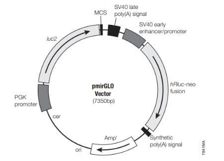
Figure 3: Schematic diagram of the pmirGLO vector
-
Cell Transfection
Transfection reagents such as PEI, calcium chloride, or Lipofectamine 2000 can be used. If using a dual-plasmid system, the ratio of the internal control plasmid to the reporter gene plasmid should be approximately 1:10 to ensure that the internal control does not interfere with the expression of the reporter gene. Perform the corresponding detection 24–36 hours post-transfection. -
Reporter Gene Detection
Lyse the cells, centrifuge to collect the supernatant, and transfer it into a 96-well plate. Add the appropriate FLuc buffer and measure the luciferase assay luminescence value. Next, add the STOP & RLuc buffer and measure the corresponding luminescence value.
-
-
Data Analysis
-
Calculate the ratio of Firefly Luciferase to Renilla Luciferase luminescence intensity for each well.
-
Calculate the average ratio of the replicate wells in the control group ($Average_{control}$). Divide the ratio of each experimental group by this average to obtain the "normalized" value, effectively setting the control group value to "1" for subsequent data analysis.
-
Plot the normalized control and experimental data using a bar chart for statistical analysis.
-
-
Application Scenarios
-
Transcription Factor-Promoter Interaction: Clone the target gene promoter into an FLuc vector, and co-transfect it with an expression vector that upregulates or inhibits the transcription factor. Changes in the FLuc signal validate its regulatory function.
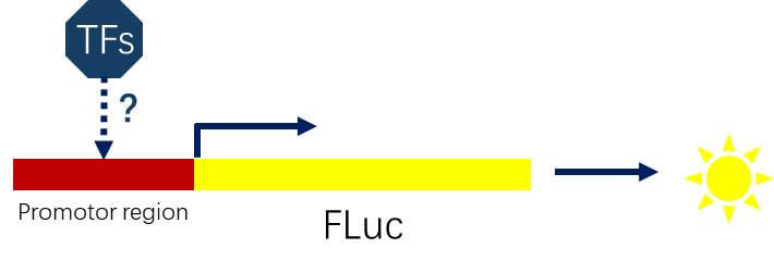
Figure 4: Diagram illustrating the study of interactions between transcription factors and target gene promoters
-
miRNA-Target Gene Verification: Insert the 3' UTR of the candidate miRNA target gene downstream of FLuc, and co-transfect it with miRNA mimics. If the miRNA binds to the target sequence, the FLuc signal decreases, demonstrating an inhibitory effect by the miRNA on that gene.
-
Signaling Pathway Activity Detection: Link a pathway response element (RE) sequence between a minimal promoter and FLuc, and co-transfect the relevant factors to build pathway reporter cell lines. Observing changes in the FLuc signal following stimulation reflects the activation state of the pathway.
-
Drug/Small Molecule Screening: Knock the Luc gene into a specific target gene using CRISPR/Cas9 to form a fusion protein. Measuring changes in the Luc signal following drug treatment enables high-throughput screening of small molecules that modulate the expression of that protein.
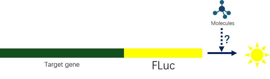
Figure 5: Diagram illustrating high-throughput screening of small molecule effects on protein expression
-
Other Applications: Luciferase assays can also be applied to the development of detection kits (e.g., combining ELISA with Luc), validating viral vector expression, and in vivo animal imaging. Due to its high sensitivity and quantifiability, the luciferase reporter assay has become the "gold standard" for gene expression and functional studies.
-
IV. Characterization and Evaluation of Luciferase Stable Cell Lines
Following the luciferase stable cell line construction, the following validations should be performed:
- Expression Level and Luminescence Stability: Measure the luminescence intensity and duration across different cell batches using a luciferase assay kit, and select clones that exhibit strong luminescence alongside slow signal decay.
- Cell Line Authentication: Perform Short Tandem Repeat (STR) profiling to confirm cell identity; concurrently, conduct mycoplasma testing to ensure the cells are contamination-free, thereby increasing experimental reproducibility.
- Functional Validation: Test relevant cellular biomarkers or phenotypes to guarantee that the integration of the Luc gene does not compromise normal cellular functions.
- Application-Based Selection: Choose an appropriate cell line source depending on research demands. For instance, Luc-labeled stable cell lines of tumor origin should be selected for in vivo tumor tracking studies, whereas easily transfectable cells like Luciferase-reported HepG2 are preferable for signaling pathway research.
V. Frequently Asked Questions (FAQ for Luciferase Assay Troubleshooting)
Q1: What should I do if the luciferase assay luminescent signal is too high (signal saturation)?
A1: When the luminescent signal exceeds the detection range of the instrument, you can reduce the amount of transfected plasmid, decrease the number of seeded cells, or appropriately dilute the cell lysate to avoid signal saturation.
Q2: What should I do if the luciferase assay signal is too low or close to background noise?
A2: If the luminescent signal is close to the background noise, you should increase the plasmid transfection amount and cell count, reduce the volume of the lysis buffer, or optimize the transfection conditions to boost the signal.
Q3: How do I handle large well-to-well variations in a dual-luciferase reporter assay?
A3: When using a multichannel pipette, ensure that each tip is securely attached and avoid generating air bubbles. For cell lysates, use the supernatant collected after centrifugation to ensure sample homogeneity and minimize well-to-well errors.
VI.Ubigene Luc Stable Cell Lines & Custom Luc Reporter Cell Line Services
As a professional cell engineering supplier, Ubigene now offers over 300 types of Luc stable cell lines, all constructed using lentiviral transduction technology to stably express Firefly Luciferase (Luc). Every cell line has undergone comprehensive luciferase activity testing, featuring strong signal specificity, high sensitivity, excellent imaging stability, and quantifiable luminescent signals.
All our Luc stable cell lines for sale are authenticated via STR profiling and verified to be free of contamination through mycoplasma testing (covering 75 common mycoplasma species). The cells are maintained at low passage numbers, possessing high viability and stable growth states, allowing them to be used directly for in vivo injection experiments. They are widely applicable to research fields such as transcription factor regulatory mechanisms, Bioluminescence Imaging (BLI), cell tracking, and efficacy evaluation.
Additionally, Ubigene provides custom services for Luc reporter cell line generation , designed to precisely monitor signaling pathway activity with high stability and reproducibility. We deliver a turn-key, one-stop service from model construction through functional validation, widely applied in drug screening, target validation, and mechanism studies. Welcome to contact us for your exclusive technical proposal and quotation! >>
 Promotions


