Expert Insights | Practical Tips for 786-0 Cell Culture and Gene Editing


The 786-O human renal clear cell adenocarcinoma cells are derived from a primary clear cell carcinoma of a 58-year-old Caucasian male. The cells possess both microvilli and desmosomes and are capable of growing in soft agar. These cells produce a PTH-like peptide that is identical to the peptides produced by breast and lung tumors. This peptide shares a similar N-terminal sequence with PTH, exhibits PTH-like activity, and has a molecular weight of 6,000 Daltons. It has been reported that these cells can form tumors in immunosuppressed hamsters. This document provides exclusive culture techniques for 786-O cells, offering a comprehensive guide to mastering 786-O cell culture and key gene editing practices.
I. Overview of Human Renal Clear Cell Adenocarcinoma Cells (786-O)
- Cell Name: Human Renal Clear Cell Adenocarcinoma Cells (786-O)
- Cell Morphology: Epithelial-like cells, adherent growth
- Culture Medium: 90% RPMI-1640 + 10% FBS
- Atmosphere: Air, 95%; Carbon dioxide, 5%
- Temperature: 37°C
- Medium Renewal Frequency: Every 2–3 days
- Passage Ratio: 1:2 – 1:4
Reference for Cell Growth Status
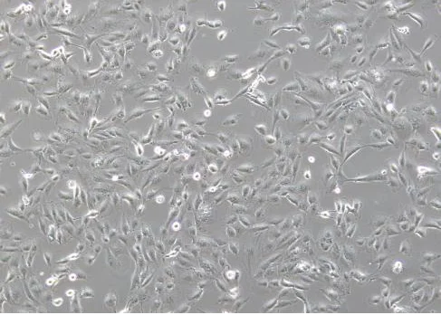
Normal Morphology: Adherent growth exhibiting epithelial-like morphology, polygonal or irregular shapes, rich cytoplasm, and may contain vacuoles. (See images below)
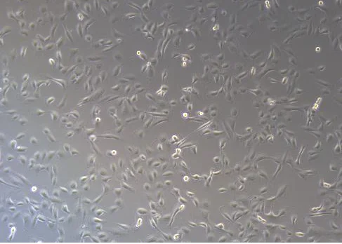
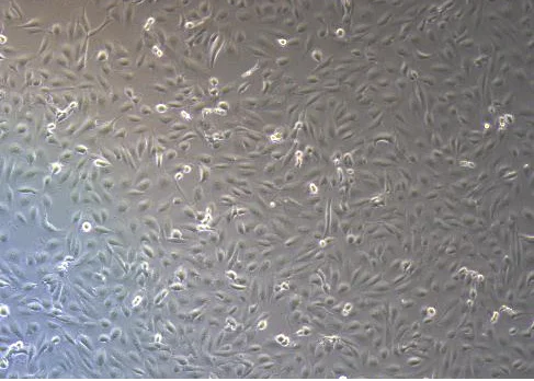
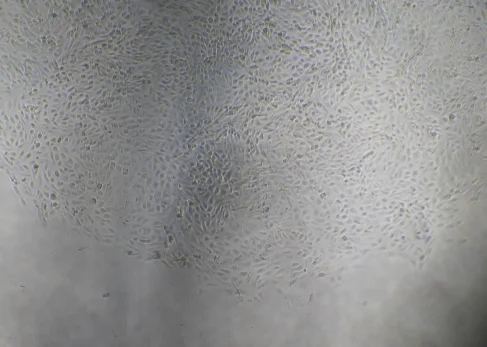
Abnormal Morphology: Cell outlines appear blurred; cells may change from spindle-shaped or polygonal to round, oval, or irregular shapes; cell volume may shrink or enlarge. (See images below)
II. 786-O Cell Thawing Procedure
-
Prepare Culture Medium
Add 7 mL of complete culture medium to a centrifuge tube and keep it ready. -
Cell Thawing
Remove the vial from dry ice. Using forceps, hold the vial cap and immerse it in a 37°C water bath. Shake gently (ensure the cap remains above the water surface). Thaw quickly for approximately 1 minute until only a small ice crystal remains. Stop water bath. -
Centrifuge Cells
Transfer the thawed cell suspension to a centrifuge tube. Centrifuge at 1100 rpm for 4 minutes and discard the supernatant. -
Resuspend and Seed Cells
Resuspend the cell pellet in complete culture medium and seed into an appropriately sized culture dish or flask. -
Culture Cells
Place the culture dish or flask in a 37°C incubator. After 24 hours, observe cell attachment and morphology.
III. 786-O Cell Subculture Procedure
-
Subculture Conditions
Subculture cells when they reach 80–90% confluency.
In a biosafety cabinet, discard the culture medium and wash the cells 1–2 times with 5 mL PBS. -
Trypsin Digestion
Add 1 mL of trypsin, gently swirl the flask to ensure the enzyme fully covers the cells.
Place the flask in a incubator for 2-3 minutes.
Under a microscope, when most cells become round and bright, gently tap the sides of the flask to detach cells, and immediately stop the digestion. -
Terminate Digestion
Add 2 mL of complete culture medium (2× the volume of trypsin) to stop the reaction.
Transfer the cell suspension to a 15 mL centrifuge tube. -
Centrifuge Cell Suspension
Centrifuge at 1100 rpm at room temperature for 4 minutes.
Discard the supernatant and resuspend the cell pellet in complete culture medium. -
Subculture and Culture
Seed cells at a split ratio of 1:2 – 1:4. A split ratio of 1:2 is recommended for the initial passage.
Observe cell morphology and attachment the next day.
IV. 786-O Cell Cryopreservation Procedure
-
Collect Cells
Harvest trypsinized cells following the standard subculture procedure and transfer them into a centrifuge tube. -
Centrifugation
Centrifuge at 1100 rpm for 4 minutes and discard the supernatant. -
Resuspend and Aliquot for Freezing
Resuspend the cell pellet in cryopreservation medium. Adjust the concentration to 1×10^6 cells/mL and aliquot 1 mL per cryovial.
Label each vial with cell line name, passage number, and date. -
Cooling and Storage
Place the vials in a controlled-rate freezing container and store overnight at -80°C.
Transfer the vials into liquid nitrogen for long-term storage.
V. Precautions for 786-O Cell Culture
-
Cell Confluency: Subculture cells at 80-90% confluency for optimal growth. For growing cells, renew with fresh complete culture medium every 2–3 days.
-
Medium Storage: Ensure the culture medium is stored at 4°C, protected from light, and used within its expiration date.
-
Strictly Adhere to Basic Culture Conditions: Ensure a normal cell culture environment and utilize the correct culture system.
-
Operational Details: Pre-warm culture medium and trypsin to 37°C prior to use to avoid thermal stress.
-
High-quality Serum: High-quality fetal bovine serum (FBS) is highly recommended for culture.
VI. Precautions for 786-O Cell Transfection
-
Cell Condition Requirements
- Ensure cells are healthy and in the logarithmic growth phase, with 70-80% confluency.
- Cell viability should be >90%, which can be assessed using Trypan Blue exclusion.
- Use low-passage cells.
- Carefully monitor trypsinization time to avoid over-digestion and cell damage.
- During the procedure, generate a single-cell suspension and avoid cell clumping.
-
Transfection Reagents and Pre-Experiment
- Mix transfection reagents thoroughly before use to ensure uniformity.
- Perform preliminary antibiotics-selection experiments to determine the optimal selection concentration post-transfection.
-
Electroporation
- Control the number of cells and seed them into appropriately sized culture plates after electroporation.
- Use gentle trypsin and terminate digestion completely with serum-containing medium.
- Wash cells 1–2 times with PBS to completely remove residual serum, preventing ionic interference with electroporation.
- Optimize electroporation parameters through preliminary experiments.
- Ensure post-electroporation cell attachment rate is ≥60%.
- Keep total electroporation time short to avoid excessive stress.
-
Lentiviral Transduction
- Perform preliminary experiments to determine the optimal MOI.
- Seed cells 18–24 hours prior to transduction; maintain cell confluency at 30–40% before virus infection, avoiding over-confluency.
- Add transduction adjuvant Polybrene prior to infection.
- Perform medium renewal 24 hours post-infection.
- Avoid repeated freezing and thawing of the viral stock used for infection.
- If transduction efficiency is too low, perform a secondary infection (ensure cells have good tolerance to the virus) or try spinoculation/centrifugal infection.
VII. Precautions for 786-O Single-Clone Seeding Experiments
-
Cell Condition Requirements:
- Use cells in the logarithmic growth phase for single-clone seeding, and the confluency prior to seeding is recommended to be controlled at around 70%.
- When cells are in optimal health, the viability for clone seeding is generally ≥90%.
-
Reagents and Pre-Experiment:
- Pre-warm all reagents (including culture medium and PBS) prior to the experiment.
- The use of gentle dissociation reagents (such as TrypLE Express) is recommended.
-
Seeding Strategy:
- Conduct preliminary experiments to determine the optimal seeding density gradient, avoiding too low a single-clone formation rate.
- When seeding 96-well plates, ensure even cell distribution. Fill the outer wells with PBS to prevent evaporation.
-
Dilution Method:
- Employ the limiting dilution method for single-clone seeding.
- After cell counting and dilution, the optimal cell count should fall within 1×10^6 to 2×10^6 cells.
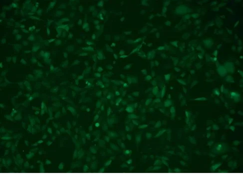
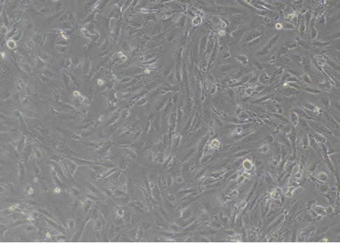
Cell Images After lentiviral infection
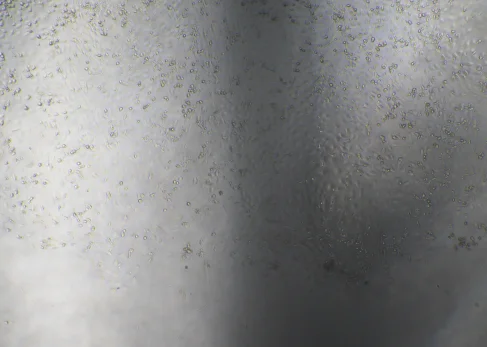
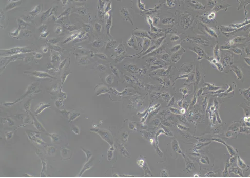
Single-Clone Cell Image
VIII. Common Issues and Solutions in 786-O Cell Culture
1. What if cell growth is slow and proliferation is poor?
Manifestation: After subculture and the same incubation period (e.g., 48 or 72 hours), the confluency is significantly lower than expected; some cells may appear shrunken, floating, or abnormal in morphology.
-
Potential Causes:
- Seeding density was too low during passage or the split ratio was too high.
- Incorrect culture conditions.
-
Solutions:
- Passage the cells when the density reaches 80%–90%. A split ratio of 1:2 is recommended for the initial passage, and subsequent ratios should be kept between 1:2 and 1:4.
- Verify that the correct culture medium (RPMI-1640 + 10% FBS) is being used.
- Confirm that the CO₂ incubator conditions are set at 37°C and 5% CO₂.
2. What if cell morphology becomes abnormal?
Manifestation: The polygonal epithelial morphology disappears, vacuoles increase, cell boundaries appear blurred, stretching/elongation occurs, and some cell debris floats in the medium.
-
Potential Causes:
- Over-digestion or overly vigorous pipetting/resuspension.
- Excessive acidity/alkalinity in pH or abnormal osmolality.
- Poor serum batch or high endotoxin levels (786-O cells are highly sensitive to serum quality).
-
Solutions:
- Properly control the digestion time; terminate immediately when cell intercellular spaces widen and cells round up under the microscope, followed by gentle pipetting (within 10 times).
- Maintain the pH between 7.2 and 7.4; HEPES solution can be supplemented if necessary.
- Serum: Use serum with low endotoxin levels and stable batch quality.
- Medium renewal can be performed every 48 hours to remove metabolic waste.
3. What if cell transfection efficiency is low?
Manifestation: Low fluorescence-positive rate and poor efficacy after antibiotic selection or treatment.
-
Potential Causes:
- Wrong selection of transfection method.
- Poor cell condition during transfection.
- Transfection parameters have not been optimized.
-
Solutions:
- When transfection efficiency remains consistently below 40%, consideration should be given to switching from liposome transfection to lentiviral transduction or electroporation.
- Ensure normal cell status prior to transfection, with cells in the logarithmic growth phase and testing negative for mycoplasma.
- Choose an appropriate seeding density according to the selected transfection method; avoid being too sparse or too dense.
- Optimize transfection parameters, such as conducting gradient preliminary experiments on nucleic acid concentration and reagent-to-nucleic-acid ratios.
Ubigene 786-O Cell Related Products Recommendations
Ubigene provides stable and reliable 786-O cells. Welcome to search our Red Cotton OmniCell Bank. All human and mouse cell lines are provided with STR authentication reports to ensure cell line identity accuracy. Ubigene offers over 1,000 wild-type cell lines covering multiple research fields, along with specialized cell culture media, to comprehensively support your scientific research needs.
786-O cell line is an ideal model for studying PTH-related diseases, such as osteoporosis. Leveraging advanced gene editing technology and platform, Ubigene has developed gene knockout and Luciferase stable cell lines based on this cell line. We also offer gene modification services for 786-O cells, including gene knockout, point mutation, gene knock-in, overexpression, and interference.
For inquiries, please contact us >>> Promotions


