
Expert Insights | Practical Tips for PANC-1 Cell Culture and Gene Editing

PANC-1 is a widely established and extensively utilized classical cell line in pancreatic cancer research. Derived from the pancreatic ductal epithelial carcinoma of a 56-year-old male Caucasian patient, the cells exhibit a typical epithelial morphology and serve as a cornerstone in vitro modeling system for pancreatic ductal adenocarcinoma (PDAC), as well as a fundamental tool for mechanistic investigations. It has been reported that this cell line harbors KRAS (Gly12Asp) and TP53 (Arg273His) driver mutations, and its proliferation can be suppressed by treatment with 1 U/mL L-asparaginase. Furthermore, PANC-1 cells display robust growth on fibroblast feeder layers and in agarose, and are capable of forming tumors in immunodeficient mice. Herein, we present a comprehensive culture protocol and a step-by-step guide to genetic manipulation in PANC-1 cells, aiming to provide researchers with a ready-to-use resource for mastering both routine maintenance and gene-editing applications in this pivotal model system.
I. Overview of Human Pancreatic Cancer Cell Line (Panc-1)
- Cell Name: Pancreatic Cancer Cell Line
- Cell Morphology: Epithelial-like, adherent
- Culture Medium: 90%DMEM+10%FBS
- Atmosphere: Air, 95%; Carbon dioxide, 5%
- Temperature: 37°C
- Medium Renewal Frequency: Every 2–3 days
- Passage Ratio: 1:2 – 1:3
Reference for Cell Growth Status:
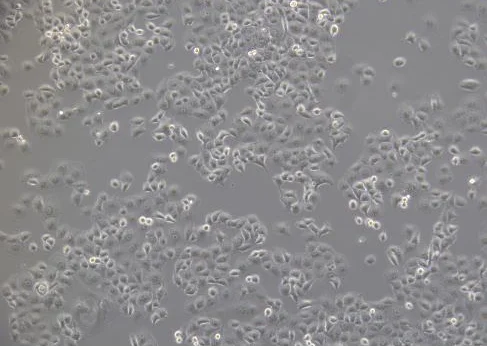
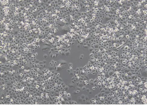
Normal morphology: Cells display an irregular polygonal shape, reminiscent of cobblestones or pebbles, and are frequently adorned with short cytoplasmic processes or spicules. The population is heterogeneous in cell size, and cultures grow as a confluent monolayer without overlapping or piling up (see figure below).
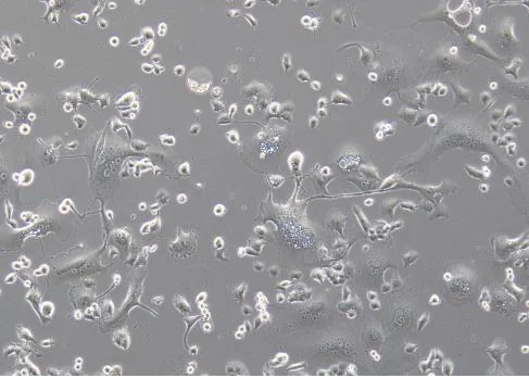
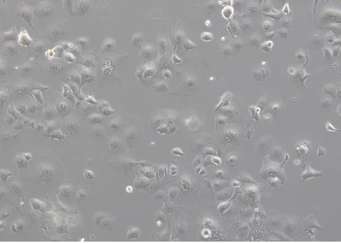
Abnormal morphology: Cells lose their characteristic polygonal architecture, exhibit pronounced enlargement in cell volume, and show increased cytoplasmic vacuolation, among other cytopathic alterations (see figure below).
II. PANC-1 Cell Subculture Procedure
- Subculture Conditions: Subculture is optimally performed when cells reach 80–90% confluence. For actively growing cultures, replace the complete medium with fresh medium every 2–3 days. When cell density exceeds 90%, cells tend to become more elongated or compressed, with a diminished polygonal morphology; however, spontaneous focal crowding or piling up is generally not observed.
- Handling details: PANC-1 cells are prone to forming aggregates. During digestion, it is essential to pipette gently but repeatedly to obtain a single-cell suspension.
- Strict culture conditions: Ensure that the cell culture environment remains optimal and that the correct culture system (medium, supplements, etc.) is employed.
- Medium storage: Store media at 4 °C away from light and use them within the indicated expiration period.
- Pre-warming: Pre-warm both the culture medium and trypsin to 37 °C prior to use to avoid temperature-induced stress.
- Serum quality: The use of high-quality foetal bovine serum (FBS) is strongly recommended for culturing this cell line.
III. Gene Editing of PANC-1 Cells
Transfection Tips
1. Cell status
- Ensure that cells are in good condition and used during the logarithmic growth phase, with confluence at 70–80%.
- Cell viability should >80%, as determined by trypan blue exclusion.
- Use cells at low passage numbers.
- Pay attention to digestion time, avoid over digestion, which may damage cells.
- This cell line tends to aggregate. During the procedure, pipette as gently as possible to obtain a single-cell suspension and minimise severe clumping.
2. Transfection reagents and preliminary experiments
- Transfection reagents must be thoroughly mixed before use to ensure homogeneity.
- It is recommended to conduct a preliminary drug screening experiment to determine the optimal concentration for antibiotic screening after transfection.
3. Electroporation
- Maintain a optimal cell number. After electroporation, seed cells into appropriate culture plates according to the cell quantity.
- Use a mild trypsin and completely neutralise digestion with serum-containing medium.
- Wash cells 1–2 times with PBS to thoroughly remove residual serum, thereby avoiding ionic interference during electroporation.
- Conduct preliminary experiments to optimise electroporation parameters.
- Ensure that the post-electroporation cell attachment rate is ≥50%.
- Keep the overall electroporation process as short as possible.
4. Lentiviral transduction
- Perform preliminary experiments to determine the optimal MOI (multiplicity of infection).
- Plate the cells 18–24 h prior to transduction; before viral infection, cell confluence should be controlled at 30–40% and should not be too high.
- Use cells at low passage numbers.
- Add the adjuvant Polybrene before infection.
- Replace the medium 24 h after infection.
- Avoid repeated freeze-thaw cycles of the viral stock used for infection.
- If transduction efficiency is too low, re-transduction can be attempted (provided that cells tolerate the virus well) or spin-transduction method can be applied.
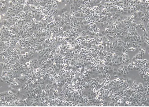
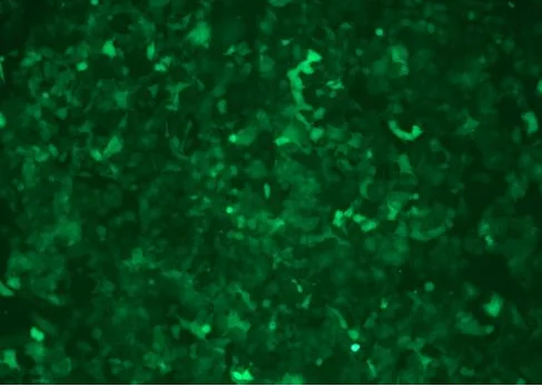
Cell morphology after lentiviral infection
Single-Cell Cloning Tips
1. Cell status
- Use cells in the logarithmic growth phase for cloning. Before plating, maintain confluence at approximately 70%.
- For cells in good condition, viability is generally ≥90% before single-cell cloning.
- Use cells at low passage numbers.
- Pay attention to digestion time, avoid over digestion, which may damage cells.
2. Reagents and preliminary experiments
- Pre-warm all reagents (including medium and PBS) in advance.
- It is recommended to use a mild digestion reagent (e.g., TrypLE™ Express).
3. Plating strategy
- Perform preliminary experiments to determine the appropriate seeding dilution gradient for single-cell cloning, avoiding low proportion of single clones.
- When seeding cells into 96-well plates, ensure even distribution; add PBS to the peripheral wells to prevent evaporation.
4. Dilution method
- Use the “limiting dilution” method for single-cell cloning.
- After counting diluted cells, the optimal cell count should fall within the range of 1 × 10⁶ to 2 × 10⁶.
IV. Common Issues and Troubleshooting in PANC-1 Cell Culture
1. Difficult digestion and severe aggregation?
Observations: After trypsinisation, cells fail to form a single-cell suspension and instead aggregate into clumps of varying sizes. Forced pipetting further compromises cell viability and morphology.
-
Possible causes:
- Cell characteristic: PANC-1 cells exhibit exceptionally strong intercellular adhesion and tight junctions. Insufficient trypsin digestion fails to disrupt these connections completely, leading to re-aggregation upon re-plating.
-
Solutions:
- Ensure complete digestion: After adding trypsin, incubate at 37 °C and monitor under the microscope. When intercellular spaces widen, cells become rounded, and edges retract, immediately neutralise the trypsin with complete medium.
- Avoid harsh pipetting: If severe clumping occurs after digestion, do not continue forceful pipetting. Instead, replate the cell clumps and allow them to attach stably for 12–24 hours; perform a second digestion and reseeding the following day.
- After seeding, avoid moving or swirling the culture vessel, as this may promote readhesion of individual cells.
2. Poor cell condition?
Observations: Abnormal morphology, extensive floating cells, retarded growth, or even cell death.
-
Possible causes:
- Bacterial, fungal, or mycoplasma contamination.
- Degradation of medium components (e.g., repeated freezethaw cycles of serum).
- Improper passaging procedures (over/under digestion, excessive centrifugation).
- Aberrant culture environment (temperature/CO₂ fluctuations).
- Cellular senescence.
-
Solutions:
- Maintain strict aseptic technique: Regularly test for mycoplasma; discard contaminated cultures immediately and thoroughly clean the incubator.
- Use highquality serum and avoid repeated freezethawing; perform regular medium changes to prevent accumulation of metabolic waste.
- Precisely control digestion time, stop trypsinisation when cells become rounded under microscopy; adjust the seeding density appropriately to avoid over/under confluence.
- Ensure stable incubator temperature, CO₂ concentration, and humidity; minimise the frequency of door openings.
3. Slow cell proliferation?
Observations: The proliferation rate is markedly slower than that of conventional tumour cell lines, resulting in prolonged passage intervals.
-
Possible causes:
- Intrinsic biological characteristics of PANC1: Its reported doubling time ranges from approximately 36 to 52 hours.
- Inoculation density is too low.
-
Solutions:
- Ensure an appropriate seeding density at the time of plating.
- Maintain patience and a rational subculture schedule: it is recommended to passage cells every 2–3 days at a split ratio of 1:2 to 1:3, when confluence reaches 80–90%.
- Perform routine medium renewal: replace the medium with fresh complete medium 2–3 times per week to sustain a favourable nutritional environment.
Ubigene PANC-1 Cell Related Product Recommendations
If you are planning to conduct research using PANC-1 cells, welcome to search for Pancreatic Cancer Cell Line (Panc-1) in Ubigene's cell bank. Ubigene offers over 1000 wild-type cell lines covering multiple research fields, and has successfully constructed stable cell lines such as gene knockout cells, Luc cells, and Cas9 cells specifically for Panc-1 cells to comprehensively meet your scientific research needs.
Contact Us for More Details>>> Promotions