
Why Does CRISPR Editing Performance Vary? Five Key Reasons Behind Differences in Editing Efficiency Across Cell Types


NHEJ and HDR are the two major DNA repair pathways responsible for CRISPR-mediated gene editing. Their activities can vary depending on the cell type. Therefore, even when the experimental system and procedures remain unchanged, an editing strategy that performs efficiently in one cell type may show little or no detectable editing in another.
These differences in editing capability among different cell lines are not always caused by problems in experimental design. Instead, they are often the result of intrinsic biological differences between cell types.
1. Cell Cycle Distribution Determines the Upper Limit of HDR Efficiency
HDR (homology-directed repair) is strictly dependent on the S/G2 phases of the cell cycle, whereas NHEJ (non-homologous end joining) can occur throughout the entire cell cycle.
For example:
Rapidly proliferating cell lines such as HEK293 and HeLa may have 30–50%
of cells in the S/G2 phase, resulting in HDR efficiencies of
approximately 10–30%.
Primary T cells and terminally differentiated neurons typically contain
less than 5% of cells in the S/G2 phase, leading to HDR efficiency
approaching zero.
Validation method:
Use flow cytometry analysis with PI/EdU staining to compare cell cycle
distributions between different cell types.
Possible solutions:
- Synchronize target cells at specific cell cycle stages (for example, using double thymidine treatment, Nocodazole synchronization, or HDR-enhancing molecules such as HDR-U+ from Yuanjing Bio);
- Switch to NHEJ-mediated knock-in strategies, such as HITI or PAGE technologies.
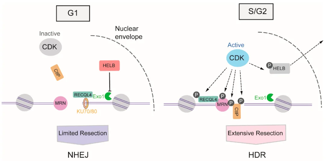
Figure 1. Cell cycle dependence of HDR
2. Differences in DNA Repair Pathway Activity Profiles: NHEJ vs HDR vs MMEJ
The double-strand breaks (DSBs) generated by Cas9 are competitively processed by the cell's endogenous DNA repair network. The expression levels of key repair factors can differ by more than ten-fold between different cell types.
Key molecules involved in repair pathways:
Core components of NHEJ: KU70/KU80, DNA-PKcs
Core components of HDR: RAD51, BRCA2, RAD52
For example:
K562 cells express high levels of KU70 but very low levels of RAD51,
causing most DNA repair events to proceed through the NHEJ pathway
rather than HDR. Some neuronal cells preferentially utilize MMEJ, which
may result in large DNA deletions.
Validation method:
Compare the expression levels of relevant proteins between two cell
types using Western blotting or qRT-PCR.
Possible solutions:
- If a cell type shows low RAD51 and high KU70 expression, HDR-based editing strategies are unlikely to succeed. Alternative approaches such as base editing (ABE/CBE) or prime editing should be considered;
- If MMEJ activity is excessively high, transient inhibition of PARP1 (for example, using olaparib) may help alter repair pathway preference.
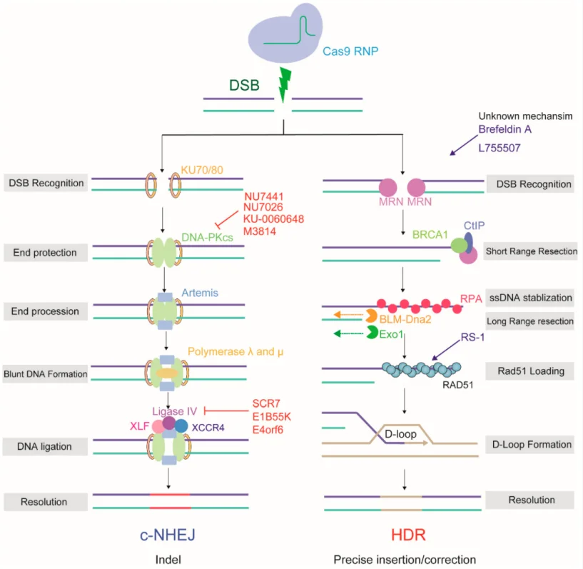
Figure 2. Key proteins required for different DNA repair pathways
3. Chromatin Accessibility: Can Cas9 Physically Access the Target DNA?
Nucleosomes represent the greatest physical barrier preventing Cas9 from binding to target DNA. Even if the PAM sequence is present, Cas9 cannot efficiently bind the DNA when the target region is packaged within condensed heterochromatin marked by modifications such as H3K9me3 and H3K27me3.
Specific histone markers:
- Regions marked by H3K4me3 and H3K27ac are typically open chromatin regions and generally show higher gene editing efficiency;
- Regions marked by H3K9me3 and H3K27me3 are associated with heterochromatin and usually exhibit lower editing efficiency.
Validation method:
Use publicly available databases (such as ENCODE ATAC-seq and Roadmap
ChromHMM) or perform ATAC-seq directly on the two cell types to compare
the accessibility of the target region.
Possible solutions:
- Before designing gRNAs, use tools such as CRISPRcut or CHOPCHOP v3, which incorporate chromatin accessibility information, to prioritize accessible regions;
- If targeting a closed chromatin region is unavoidable, consider pretreatment with epigenetic-modifying drugs.
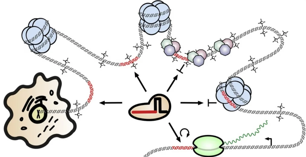
Figure 3. Heterochromatin regions may impact editing efficiency
4. Cell-Type-Dependent Differences in Delivery Efficiency
Different cell types show substantial differences in their tolerance and intrinsic efficiency for physical transfection, chemical transfection, and viral delivery methods.
Common scenarios:
- Electroporation: Primary T cells and iPSCs often show survival rates below 50% after electroporation. The proportion of DNA/RNP molecules successfully entering the nucleus is also lower.
- Lipid-based transfection: Suspension cells such as Jurkat and K562 generally show transfection efficiencies below 20%. Adherent cells such as HEK293 can achieve efficiencies above 80%.
- Lentiviral delivery: Integration efficiency is affected by the expression levels of cellular surface receptors, such as CD4 and LDLR.
Validation method:
Treat two cell types simultaneously with a GFP expression plasmid or
fluorescently labeled Cas9 RNP, then use flow cytometry to quantify: The
percentage of positive cells; Fluorescence intensity levels.
Possible solutions:
- For difficult-to-transfect cells, prioritize lentiviral delivery approaches;
- Optimize electroporation parameters and add apoptosis inhibitors when necessary;
- Consider nanoparticle-based delivery methods, such as lipid nanoparticles (LNPs).
5. Target Sequence Polymorphisms: SNPs and PAM Mutations
Even when targeting the same gene, different cell lines or donor-derived cells may contain single nucleotide polymorphisms (SNPs) within the gRNA target region. This is particularly important because nucleotide mismatches located within 10 bp of the PAM sequence can reduce Cas9 cleavage efficiency by more than 90%.
Hidden risks:
- A G→A mutation within the PAM sequence (for example, the NGG recognition site of SpCas9) can completely eliminate Cas9 recognition.
- SNPs within the seed region (the 8–12 nt sequence at the 3′ end of the gRNA) have the greatest impact.
- Even synonymous mutations or intronic mutations can affect editing outcomes.
Validation method:
Amplify the target region from both cell types using PCR, followed by
Sanger sequencing, and compare sequence differences. Note: Reference
sequences in cell bank databases may sometimes differ from the actual
experimental cell lines.
Possible solutions:
- During gRNA design, prioritize highly conserved genomic regions based on cross-species sequence alignment;
- Re-sequence the target region in the specific cell line before starting experiments;
- If SNPs are identified, redesign the gRNA to account for the variant sequence and utilize mismatch tolerance.
6. Conclusion
Therefore, when performing CRISPR gene editing, a strategy validated successfully in one cell type should not simply be transferred directly to another cell type. In many cases, preliminary experiments are required to rapidly evaluate editing performance before deciding whether to maintain the original strategy or switch to a different technical approach.
Only by developing editing strategies tailored to the characteristics of each cell type can the full potential of the CRISPR-Cas9 system be achieved.
EZ-HRex™ Technology by Ubigene Biosciences
EZ-HRex™ is a proprietary CRISPR/Cas9-based gene editing technology developed by Ubigene Biosciences for efficient generation of gene point mutation cell models. Compared with conventional approaches, EZ-HRex™ introduces the innovative HDR-U+ molecule. Following transfection, the proportion of HDR genotypes at the Cell Pool level can reach up to 84%. This technology integrates multiple core capabilities, including:
- Effective regulation of the cell cycle to promote more cells entering the S/G2 phase after transfection;
- A unique gRNA design algorithm based on cellular genomic characteristics;
- Optimization strategies developed through extensive exploration of gene editing parameters across thousands of cell lines;
- Accurate detection methods for Cell Pool editing efficiency;
- Improved monoclonal formation strategies and high-throughput identification methods for low-frequency genotypes.
Ubigene Biosciences has successfully achieved gene editing in more than 500 cell types and completed over 10,000 gene editing projects, providing efficient cell gene editing services for researchers worldwide.
Contact us for more information! Promotions