
The OPA1-NRF1 Axis direct dendritic cell function in antitumor immunity


Introduction
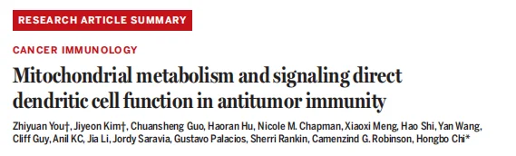
Conventional type 1 dendritic cells (cDC1s) are critical for orchestrating CD8⁺ T cell-mediated antitumor immunity. However, they are susceptible to metabolic stress and functional impairment within the tumor microenvironment, and the mechanisms by which mitochondrial metabolism regulates their function remain largely elusive. A team led by Hongbo Chi at St. Jude Children‘s Research Hospital published a study in Science , revealing that intratumoral cDC1s exist in two distinct mitochondrial states. The OPA1-NRF1 signaling axis governs cDC1 antigen presentation and antitumor function by sustaining mitochondrial oxidative phosphorylation (OXPHOS), energy metabolism, and redox balance. Moreover, adoptive transfer of cDC1s with polarized mitochondria combined with immune checkpoint blockade (ICB) markedly enhanced antitumor efficacy.
Background
cDC1s are essential antigen-presenting cells that drive CD8⁺ T cell activation and determine the outcome of ICB therapy. However, they often exhibit metabolic dysregulation and functional decline within the tumor microenvironment. Previous studies have suggested that mitochondrial OXPHOS is dispensable for dendritic cell maturation and may even be associated with immune tolerance. Thus, how mitochondrial metabolism regulates cDC1 antitumor function in the tumor microenvironment remains a key unresolved question.
Objectives
To characterize the heterogeneity of mitochondrial states in intratumoral cDC1s, elucidate the molecular mechanisms by which OPA1-mediated mitochondrial metabolic signaling governs cDC1 antitumor immunity, and validate the feasibility of targeting this pathway to optimize cancer immunotherapy.
Methods
- Animal models and gene editing: Generation of DC-specific OPA1/DRP1 knockout, cDC1-specific OPA1 knockout, and other mouse models; establishment of multiple syngeneic tumor models.
- Cellular analysis: Flow cytometric sorting of cDC1s; measurement of mitochondrial membrane potential and mass to define functional subpopulations.
- Functional assays: Antigen presentation assays, Seahorse mitochondrial respiration analysis, and immunoblotting.
- Multi-omics profiling: Proteomics, metabolomics, ATAC-seq, and transcriptomic analyses.
- Therapeutic validation: Adoptive transfer of [TMRM/MG]hi or [TMRM/MG]lo cDC1s, in combination with ICB, to evaluate antitumor efficacy.
Workflow
- Clinical/in vivo discovery: Identification of two distinct mitochondrial states in human and mouse intratumoral cDC1s.
- Key factor identification: OPA1 orchestrates mitochondrial morphology and antitumor function in cDC1s.
- Mechanism: OPA1 sustains OXPHOS via NRF1, thereby suppressing autophagy and maintaining redox balance.
- Dynamic changes: Progressive mitochondrial dysfunction in cDC1s during tumor progression.
- Rescue validation: DRP1 deletion restores the functional defects caused by OPA1 deficiency.
- Translational application: Polarized cDC1s combined with ICB achieve potent antitumor immunity.
Key Findings
1. OPA1 orchestrates mitochondrial states and antitumor CD8⁺ T cell responses in cDC1s
Intratumoral cDC1s comprised two subpopulations ([TMRM/MG]hi and [TMRM/MG]lo), with the former exhibiting superior capacity to prime CD8⁺ T cells. OPA1 is selectively upregulated in cDC1s. DC-specific OPA1 deletion resulted in mitochondrial fragmentation and impaired antitumor immunity.
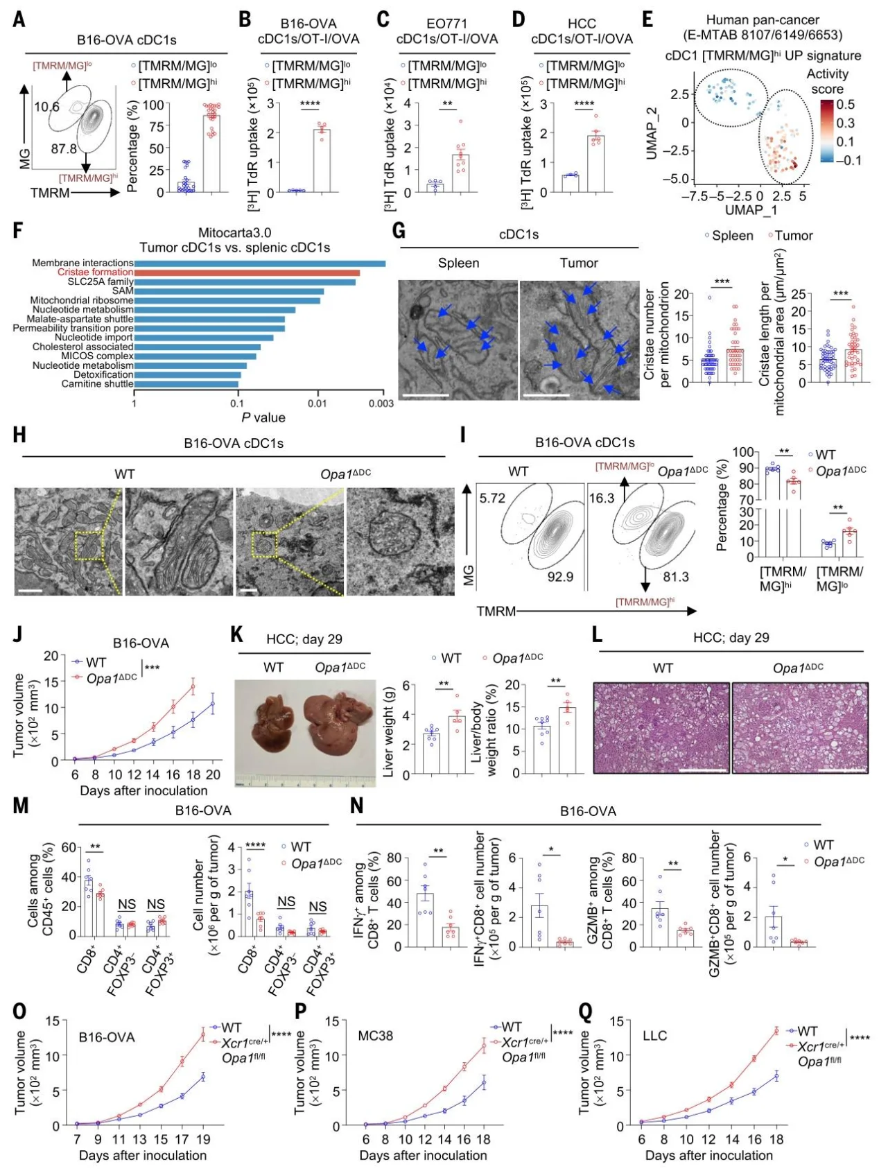
Fig. 1. OPA1 orchestrates discrete mitochondrial states of cDC1s and their function in activation of antitumor CD8+ T cell responses.
2. The OPA1-NRF1 axis-mediated OXPHOS promotes cDC1 antigen presentation
OPA1 deficiency selectively reduced surface MHC-I-antigen complex levels and NRF1 transcriptional activity. ETC integrity is essential for cDC1 function, with OXPHOS serving as the core downstream effector.
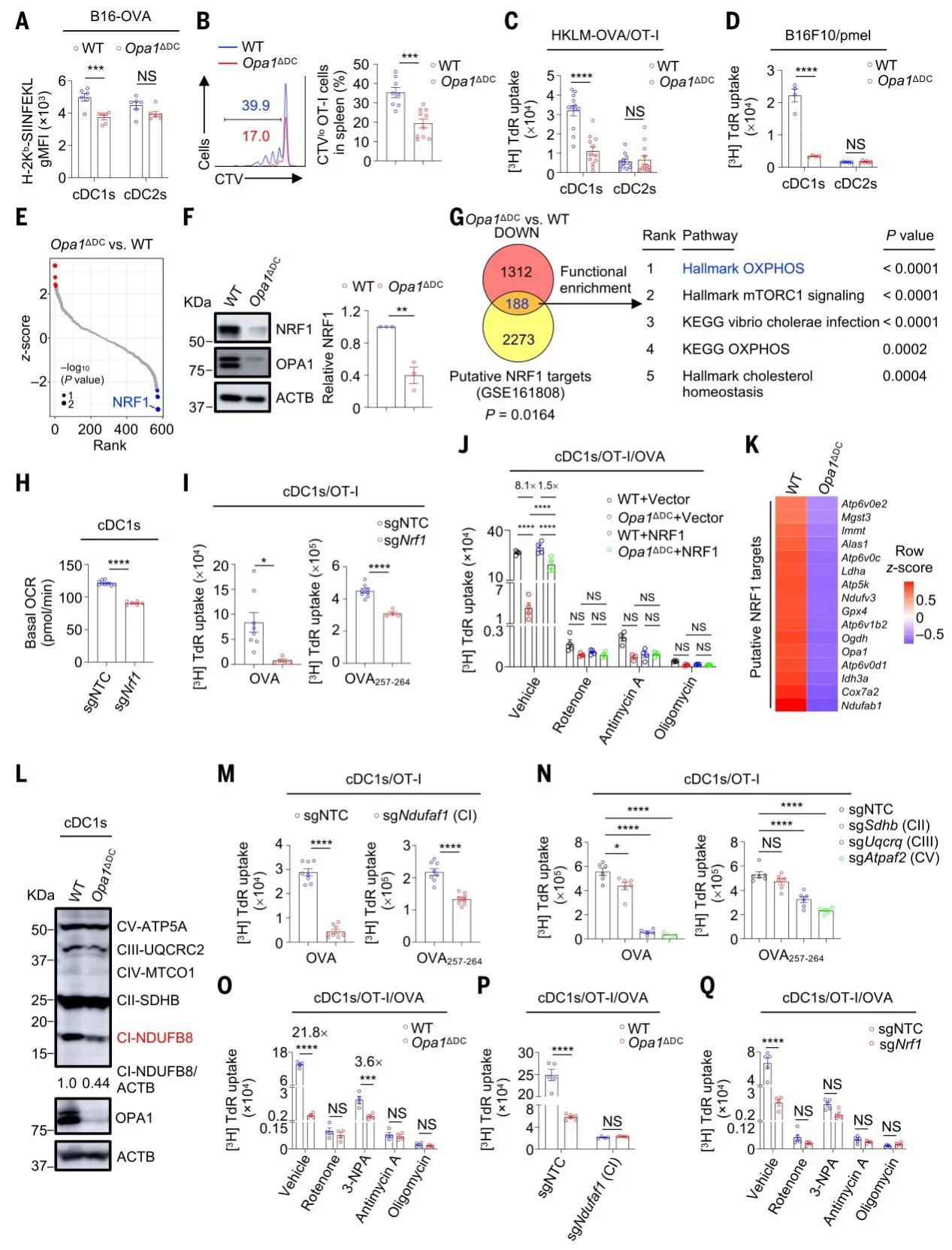
Fig. 2. OPA1-NRF1 axis–mediated OXPHOS promotes cDC1 antigen presentation.
3. Mitochondrial OXPHOS suppresses autophagy and maintains NAD⁺ balance
OPA1/NRF1 deficiency led to decreased ATP, AMPK activation, and upregulated autophagy, causing MHC-I degradation. DRP1 deletion reversed these effects, confirming the role of the mitochondrial fusion-fission balance.
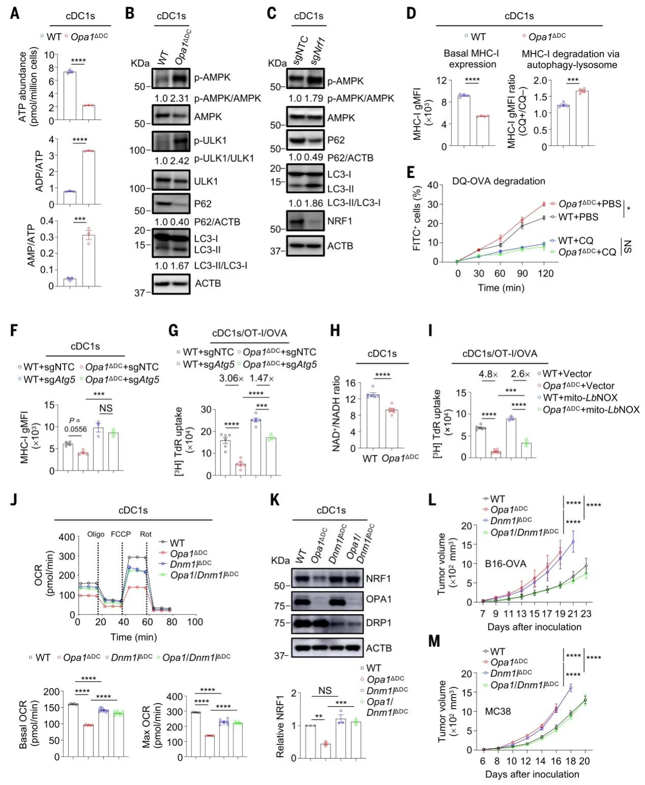
Fig. 3. Mitochondrial OXPHOS–driven autophagy inhibition and NAD+ regeneration contribute to OPA1-mediated cDC1 functional fitness.
4. Progressive mitochondrial dysfunction in cDC1s
Intratumoral cDC1 mitochondrial fitness declines during tumor progression. Adoptive transfer of [TMRM/MG]hi cDC1s combined with ICB achieved complete tumor regression.
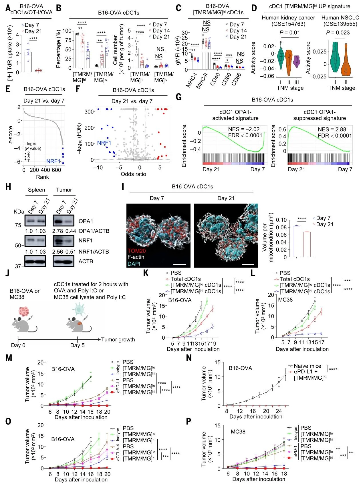
Fig. 4. Intratumoral cDC1s experience a progressive decline of mitochondrial membrane potential and volume during tumor progression, which is associated with down-regulation of OPA1 and NRF1 levels.
Significance and Innovations
- Conceptual advance: Revealed mitochondrial state heterogeneity and the OPA1-NRF1-OXPHOS axis.
- Mechanistic breakthrough: Uncovered how OXPHOS sustains MHC-I stability via autophagy suppression.
- Translational potential: Proposed a strategy combining polarized cDC1s with ICB.
- Actionable targets: Identified OPA1, NRF1, and DRP1 as core targets for metabolic engineering.
Conclusion
This study demonstrates that the OPA1-NRF1 axis safeguards cDC1 function by sustaining OXPHOS and inhibiting autophagy-mediated MHC-I degradation. These findings provide a new framework for metabolically engineering dendritic cells to optimize cancer immunotherapy.
Ubigene Biosciences remains committed to "Making Gene Editing Easier," with over 13,000 successful cases and 10,000+ KO cell products. Our technology enhances editing efficiency by 10-20X, serving over 10,000 laboratories and pharmaceutical companies.
OPA1 plays an essential role in regulating mitochondrial fusion, critical for morphology and function. Mitochondria are involved in apoptosis and calcium homeostasis. If you are interested in OPA1 research, Ubigene offers OPA1 gene knockout services.
Welcome to contact us for your exclusive technical proposal and quotation! >> Promotions