
What Are Reporter Gene Cell Lines? An Overview of Standardized “Biosensors” for Studying Gene Expression Regulation


I. Introduction
Reporter genes technology is an essential tool in gene expression studies, enabling the visualization of intracellular transcriptional regulatory events through measurable reporter protein signals. Reporter genes encode easily detectable enzymes or proteins and are commonly inserted downstream of regulatory elements such as promoters and enhancers. Due to their rapid and highly sensitive detection capabilities, reporter systems allow experimental results to be obtained within a short period of time and are widely applied in high-throughput screening applications. Commonly used reporter genes include luciferase, fluorescent proteins (e.g., EGFP), and β-galactosidase . Among these, firefly luciferase, derived from the North American firefly (Photinus pyralis), catalyzes the oxidation of luciferin to generate bioluminescence. Luciferase-based assays have become one of the most widely used reporter systems due to their non-radioactive detection format, high sensitivity, rapid response kinetics, relatively long half-life, and extremely low background signal in host cells.
In biopharmaceutical quality control and drug discovery, reporter cell lines are extensively utilized for signaling pathway activity analysis, target validation, and compound screening. Compared with conventional detection approaches, reporter cell lines enable dynamic and quantitative monitoring of pathway activation, offering advantages including high accuracy and excellent reproducibility. Establishing stably expressing reporter cell lines is therefore critical for ensuring assay consistency and comparability. In recent years, the emergence of CRISPR/Cas9-mediated targeted integration technology has enabled precise insertion of exogenous reporter genes, further accelerating advances in this field.
II. Key Technical Principles
The fundamental principle of the firefly luciferase reporter system is illustrated in Figure 1. A promoter containing specific pathway-responsive elements (such as NF-κB binding sequences) is engineered into a plasmid carrying the Luc gene. Following delivery into host cells, the construct is integrated to establish a stable reporter cell line. When an external stimulus activates the corresponding signaling pathway, activated transcription factors bind to the specific response elements and drive transcription of the Luc gene, resulting in luciferase protein expression. Upon addition of the substrate luciferin, luciferase catalyzes its oxidation reaction to produce a luminescent signal. The intensity of emitted light is proportional to the activity of the corresponding signaling pathway.
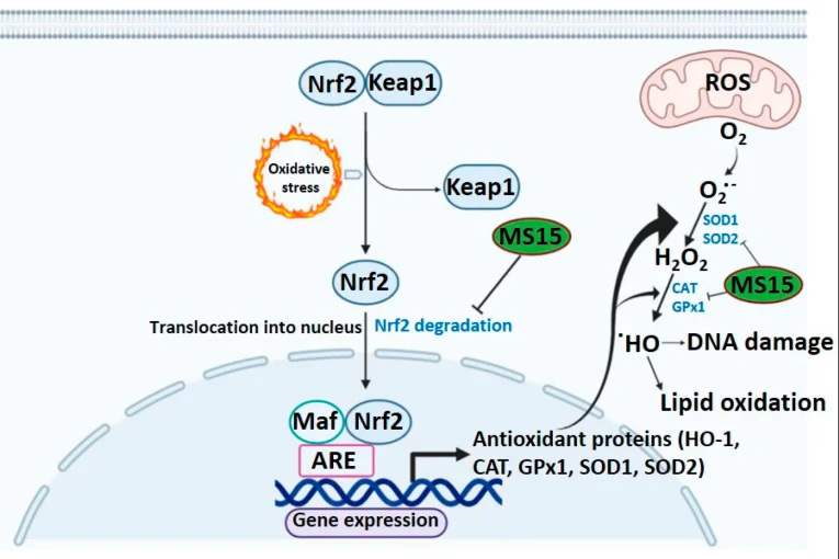
Figure 1. Schematic overview of the firefly luciferase reporter system
In this system, cellular stimulation (such as treatment with TNF-α or small-molecule compounds) activates intracellular signaling cascades, ultimately inducing Luc gene expression and generating a luminescent output. The dual-luciferase system is commonly used for internal normalization: one reporter is the luciferase of interest (e.g., firefly luciferase), while the other is a Renilla luciferase reporter as an internal control for transfection efficiency and cell viability. The major advantages of luciferase-based reporter systems include highly specific enzyme-substrate reactions that minimize autofluorescence interference, exceptional sensitivity, extremely low background signals, and compatibility with live-cell or in vivo imaging applications. Firefly luciferase produces yellow-green bioluminescence with relatively favorable tissue penetration properties, making it suitable for in vivo tracking. In comparison, fluorescent proteins require external excitation light and are more susceptible to background interference. They are therefore generally preferred for live-cell localization and imaging, rather than highly sensitive quantitative measurements.
III. Construction Strategies and Workflow for Luciferase Reporter Cell Lines
Key steps in constructing luciferase reporter cell lines include: vector design, cell transfection/transduction, positive clone selection, and functional validation. Detailed strategies are as follows:
- Selection of Response Element and Promoter : The first step is to select appropriate pathway-responsive elements (e.g., NF-κB RE, ARE, etc.) according to the research objective. These regulatory sequences are typically arranged as multiple tandem repeats to enhance signal amplification and are positioned upstream of minimal promoters (e.g., SV40, TK, or promoterless plasmids such as pGL4.20). For systems that do not require pathway-specific responses, constitutive strong promoters (e.g., CMV, EF1α) can be used to directly drive luciferase expression for in vivo imaging or cell tracking applications.
-
Selection of Reporter Gene Variants:
Firefly luciferase is one of the most commonly used reporter genes,
utilizing D-luciferin as its substrate and producing light emission
at approximately 560 nm. Renilla luciferase uses coelenterazine as
its substrate and emits light at approximately 480 nm. Due to its
distinct spectral properties, Renilla luciferase can be combined
with firefly luciferase in dual-reporter assays for normalization
and comparative analysis. NanoLuc luciferase is a next-generation,
ultra-bright luciferase variant with substantially higher
luminescence intensity than conventional luciferases. It emits
shorter-wavelength blue light (~460 nm), making it particularly
suitable for dual-reporter assays or applications requiring
extremely high sensitivity.
For applications requiring secreted reporter proteins, Gaussia luciferase or Metridia luciferase can be selected. Reporter constructs can be generated using commercially available plasmids (such as Promega dual-luciferase vectors) or custom-designed expression vectors.
For in vivo imaging applications, engineered luciferases with red-shifted emission spectra can be considered to improve tissue penetration and imaging performance. Dual-reporter systems (such as Firefly luciferase + Renilla luciferase) allow simultaneous measurement of two signals. Typically, the experimental reporter gene and the internal control reporter are either incorporated into the same vector or introduced through co-transfection of two independent vectors.
-
Vector Construction and Cell Transfection/Transduction:
Clone the designed response element–promoter fragment into the
reporter vector to generate the luciferase expression plasmid. The
plasmid can be introduced into target cells through
lentiviral-mediated transduction. Lentiviral transduction generally
provides higher delivery efficiency, particularly in
difficult-to-transfect cell types, and facilitates stable genomic
integration of the reporter construct. During transfection or
transduction experiments, appropriate controls, including
empty-vector controls and positive controls, should be included to
evaluate reporter activity and experimental reliability.
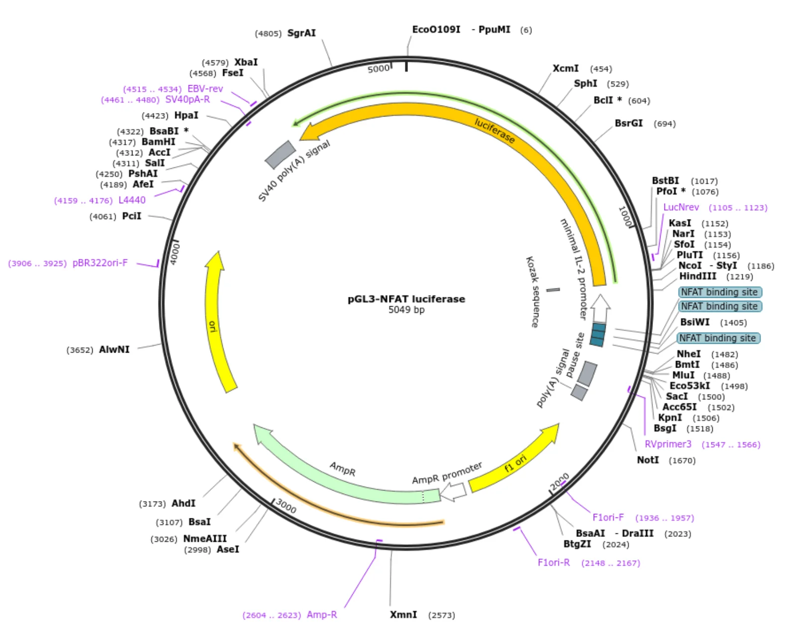
Figure 2. Vector backbone
- Selection and Clonal Isolation: Reporter vectors generally contain resistance markers, such as neomycin or puromycin resistance genes. Following transfection or transduction, cells are subjected to antibiotic selection using an appropriate concentration of selection reagent. After obtaining a stably expressing polyclonal cell population, single-cell cloning can be performed to generate monoclonal reporter cell lines. Common approaches include limiting dilution cloning or fluorescence-activated cell sorting (FACS)-based single-cell isolation. Each individual clone should be evaluated for reporter activity, typically through luminescence intensity measurement, to identify clones exhibiting strong and stable reporter expression.
IV. Data Analysis and Interpretation
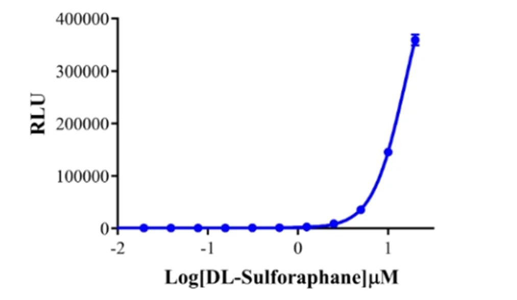
The primary output of reporter gene assays is the luminescence or fluorescence intensity generated by reporter-expressing cells. Conventional data analysis typically involves generating concentration-response curves for pathway agonists or inhibitors, calculating pharmacological parameters such as IC50 values, and performing statistical analysis to evaluate experimental significance. For example, DL-Sulforaphane exhibits a typical dose-dependent activation effect in ARE-Luc (Nrf2 antioxidant pathway) HEK293 reporter cells: as compound concentration increases, luciferase signal (RLU) remains at baseline at low concentrations, rises sharply after a certain threshold, and plateaus at high concentrations, presenting a classic sigmoidal dose-response curve. This indicates that DL-Sulforaphane significantly enhances downstream transcriptional activity through activation of the Keap1-Nrf2-ARE signaling axis. This system offers a favorable dynamic range and sensitivity, making it suitable for functional evaluation and screening of Nrf2 pathway agonists.
V. Applications and Limitations
Luciferase reporter cell lines have been widely applied in signaling pathway analysis, target validation, drug discovery, and bioactivity assessment of biopharmaceutical products. For example, these systems can be used to identify activators or inhibitors of signaling pathways including NF-κB, Wnt/β-catenin, p53, and GPCR signaling, as well as to evaluate receptor-ligand interactions. Because bioluminescent signals can be monitored dynamically over time, reporter cell lines can also be applied in live-cell imaging and in vivo tracking studies when combined with appropriate imaging platforms and reporter constructs.
However, reporter gene cell lines have certain limitations: First, each cell line is specifically responsive to a particular pathway or element and cannot be universally applied to all pathways. Establishing cell lines for new pathways is time-consuming and resource-intensive. Second, exogenous expression of reporter proteins may affect host cell function, necessitating validation that cell growth and basic phenotypes remain normal. Additionally, luciferase-based reporter assays provide an indirect measurement of pathway activity and cannot directly reflect downstream molecular events such as protein post-translational modifications or protein activation states. Certain compounds may directly inhibit luciferase enzymatic activity rather than the pathway itself, so appropriate control experiments are required to exclude potential false positives. Finally, luciferase assays require substrate addition and plate-reader measurement. Compared with fluorescent protein-based approaches, luciferase assays generally involve additional experimental steps.
| Reporter System / Tool | Advantages | Limitations | Typical Applications |
| Firefly Luciferase (Firefly Luc) | High sensitivity; broad linear detection range; strong luminescent signal; minimal background interference due to negligible endogenous expression in mammalian cells | Requires exogenous substrate (luciferin); ATP-dependent; relatively long half-life; assay conditions require strict temperature control | Gene expression analysis; drug screening; in vitro and in vivo imaging |
| Renilla Luciferase (Renilla Luc) | Rapid reaction kinetics; ATP-independent; commonly used as an internal control due to distinct emission spectrum from Firefly luciferase | Lower luminescence intensity compared with Firefly Luc; requires co-elenterazine substrate; shorter half-life requiring rapid signal detection | Dual-luciferase normalization assays; transcription factor activity studies |
| NanoLuc Luciferase (NanoLuc) | Extremely high luminescence intensity; stable and persistent signal; ATP-independent | Requires specialized substrate (furimazine); short-half-life variants require rapid measurement; different emission spectrum from Firefly luciferase | Highly sensitive quantitative assays; monitoring of cell proliferation and dynamic signaling changes |
| β-Galactosidase (β-Gal) | Stable enzymatic activity; simple detection; does not require ATP | Requires cell lysis and chemical colorimetric detection; slow reaction kinetics; relatively high background noise; unsuitable for dynamic live-cell monitoring | Conventional reporter assays; gene cloning screening |
| Fluorescent Proteins (e.g., EGFP) | No requirement for exogenous substrates; direct visualization by fluorescence microscopy; suitable for live-cell imaging; relatively long half-life | Requires excitation light; susceptible to autofluorescence interference from tissues or cells; photobleaching; relatively lower sensitivity | Live-cell localization and dynamic cellular imaging |
| Dual-Luciferase System (Firefly + Renilla) | Enables simultaneous measurement of experimental reporter and internal control signals; normalizes transfection variability and improves quantitative accuracy | More complex workflow requiring two detection reagents; relatively higher assay cost | High-throughput drug screening; quantitative transcription factor analysis; evaluation of cellular toxicity effects |
VI. Ubigene Luc Reporter Cell Line Services
Ubigene provides customized luciferase (Luc) reporter cell line development services based on established molecular cloning technologies and stable cell line engineering platforms. According to specific research requirements, customized reporter systems can be developed for key signaling pathways, including Nrf2/ARE, NF-κB, MAPK, and PI3K-AKT pathways, enabling highly sensitive and real-time quantitative monitoring of intracellular signaling activity changes. The constructed reporter cell models are primarily generated through lentivirus-mediated stable integration strategies, providing excellent genetic stability, batch-to-batch consistency, and high signal-to-noise ratios. These advantages effectively minimize background interference, improve data reproducibility, and enhance experimental reliability. These systems are well suited for drug screening, mechanism-of-action studies, and high-throughput compound evaluation across a variety of application scenarios. Contact us to learn more >>>
 Promotions