
IF=21.8 | Ubigene EGFP cells Empower Magnetic Responsive Lipid nanoparticles for Rapid mRNA Delivery with over 33-Fold Higher In Vivo Efficiency

Introduction
Conventional mRNA delivery systems often require several hours to achieve efficient cellular internalization, during which non-internalized mRNA payloads are gradually degraded. A research team led by Professor Bin Li from Southern University of Science and Technology recently published a study in Advanced Composites and Hybrid Materials (IF = 21.8), reporting a magnetic responsive lipid nanoparticle (MrLNP) platform capable of rapid mRNA delivery under external magnetic stimulation. In vivo delivery efficiency was improved by more than 33-fold compared with conventional non-magnetic LNPs, providing a promising strategy for acute and critical diseases that require immediate protein expression. In this study, Ubigene supplied stable EGFP-expressing human embryonic kidney cells (293T-EGFP) as a quantitative and visualizable model for evaluating genome editing efficiency. These cells were used to validate the co-delivery of CRISPR-Cas9 mRNA and sgRNA mediated by MrLNPs, as well as the resulting functional gene knockout efficiency.
Research Background
mRNA-based therapeutics possess promising clinical potential due to their high designability and lack of genomic integration risk. However, safe and efficient intracellular delivery remains one of the major bottlenecks limiting broader clinical translation.
Lipid nanoparticles (LNPs) are currently the leading non-viral delivery vehicles for mRNA therapeutics and have already been successfully applied in COVID-19 mRNA vaccines. Despite their success, conventional LNP-mediated mRNA internalization is relatively slow, which compromises delivery efficiency. Magnetic nanoparticles (MNPs), by contrast, possess magnetic-responsive properties that enable targeted and accelerated drug delivery under external magnetic fields. While MNPs have demonstrated broad potential in drug delivery applications, their use for in vivo mRNA delivery has remained largely unexplored.
Research Objectives
Construct magnetic responsive lipid nanoparticles (MrLNPs) and characterize their physicochemical and magnetic-responsive properties; Demonstrate the broad applicability of MrLNPs across multiple mRNA payloads and diverse cell types; Evaluate the efficiency, protein expression capability, and biosafety of rapid mRNA delivery mediated by MrLNPs both in vitro and in vivo; Investigate the intracellular trafficking mechanisms and translational potential of magnetically accelerated mRNA uptake.
Research Workflow
- A novel ionizable lipid, CF3-3N6-UC18, was synthesized and formulated into mRNA-loaded LNPs. Oligo-coupled magnetic beads were then electrostatically adsorbed onto the nanoparticle surface to construct MrLNPs.
- Key preparation parameters, including the mRNA-to-bead ratio, mixing method, and post-mixing resting time, were systematically optimized.
- The particle size, zeta potential, morphology, and magnetic responsiveness of MrLNPs were characterized, and their magnetic-guided delivery and cell sorting capabilities were validated.
- The in vitro delivery performance of MrLNPs was evaluated across multiple mRNA payloads, cell lines, and 3D cell culture systems, including assessments of genome editing and gene silencing efficiency.
- Cellular uptake pathways, endosomal escape behavior, and in vitro biocompatibility of MrLNPs were investigated.
- In vivo mRNA delivery efficiency, biodistribution, and biosafety of MrLNPs under magnetic stimulation were further evaluated.
Key Findings
1. Preparation, Optimization, and Characterization of CF3-3N6-UC18 MrLNPs
The researchers first synthesized a novel ionizable lipid, CF3-3N6-UC18, which exhibited an order-of-magnitude improvement in in vivo mRNA delivery efficiency compared with the previously reported CF3-2N6-UC18 formulation. The chemical structure was confirmed by high-resolution mass spectrometry (HRMS) and nuclear magnetic resonance (NMR) spectroscopy.
mRNA was subsequently encapsulated into CF3-3N6-UC18 LNPs, followed by electrostatic adsorption of oligo(dT)25 magnetic beads onto the nanoparticle surface, successfully generating magnetic responsive lipid nanoparticles (MrLNPs).
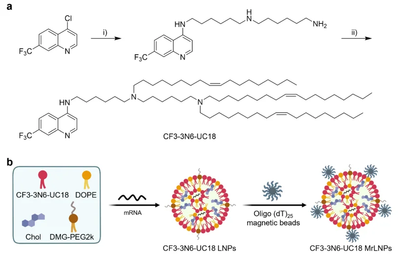
Figure 1. Construction of CF3-3N6-UC18 MrLNPs for mRNA Delivery
Using mRNA transfection efficiency as the evaluation metric, the researchers systematically optimized the mRNA-to-bead weight ratio, mixing method, and post-mixing resting time in a stepwise manner. The results identified an mRNA-to-bead weight ratio of 1:25, vigorous pipette mixing, and a 5-minute resting period as the optimal preparation conditions. Under these parameters, MrLNPs enabled rapid mRNA delivery in the presence of an external magnetic field, whereas delivery efficiency in the absence of magnetic stimulation showed no significant difference compared with conventional LNPs.
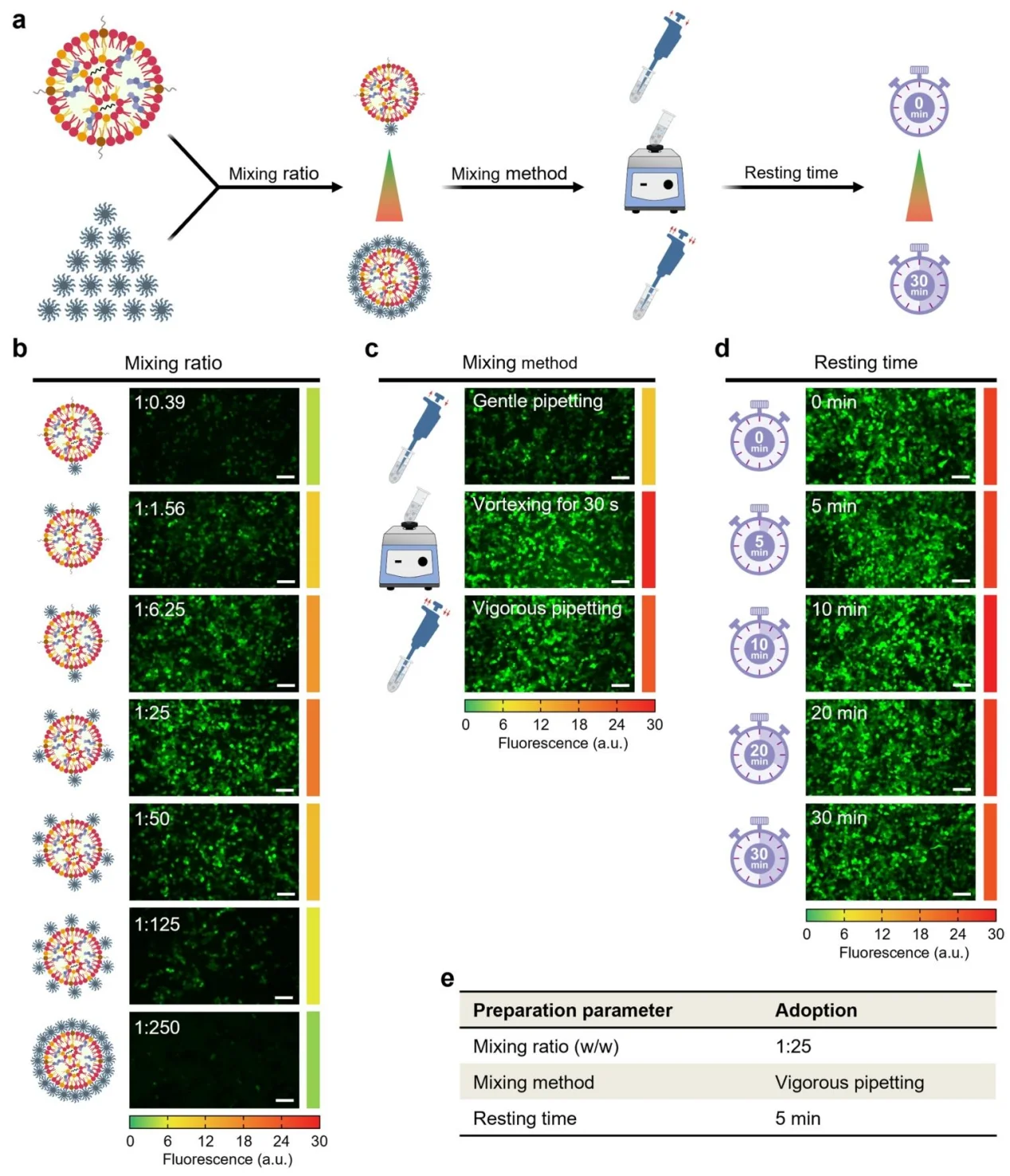
Figure 2. Optimization of MrLNP Preparation Parameters
Dynamic light scattering (DLS) and scanning electron microscopy (SEM) analyses revealed that magnetic beads tended to aggregate on their own, while dispersion improved substantially after association with LNPs. Zeta potential measurements confirmed electrostatic assembly between positively charged LNPs and negatively charged magnetic beads, with MrLNPs exhibiting a zeta potential of 25.1 mV.
Agarose gel electrophoresis further demonstrated that MrLNPs effectively encapsulated mRNA payloads while retaining magnetic responsiveness, enabling magnetically guided selective cell delivery and sorting of transfected cells.
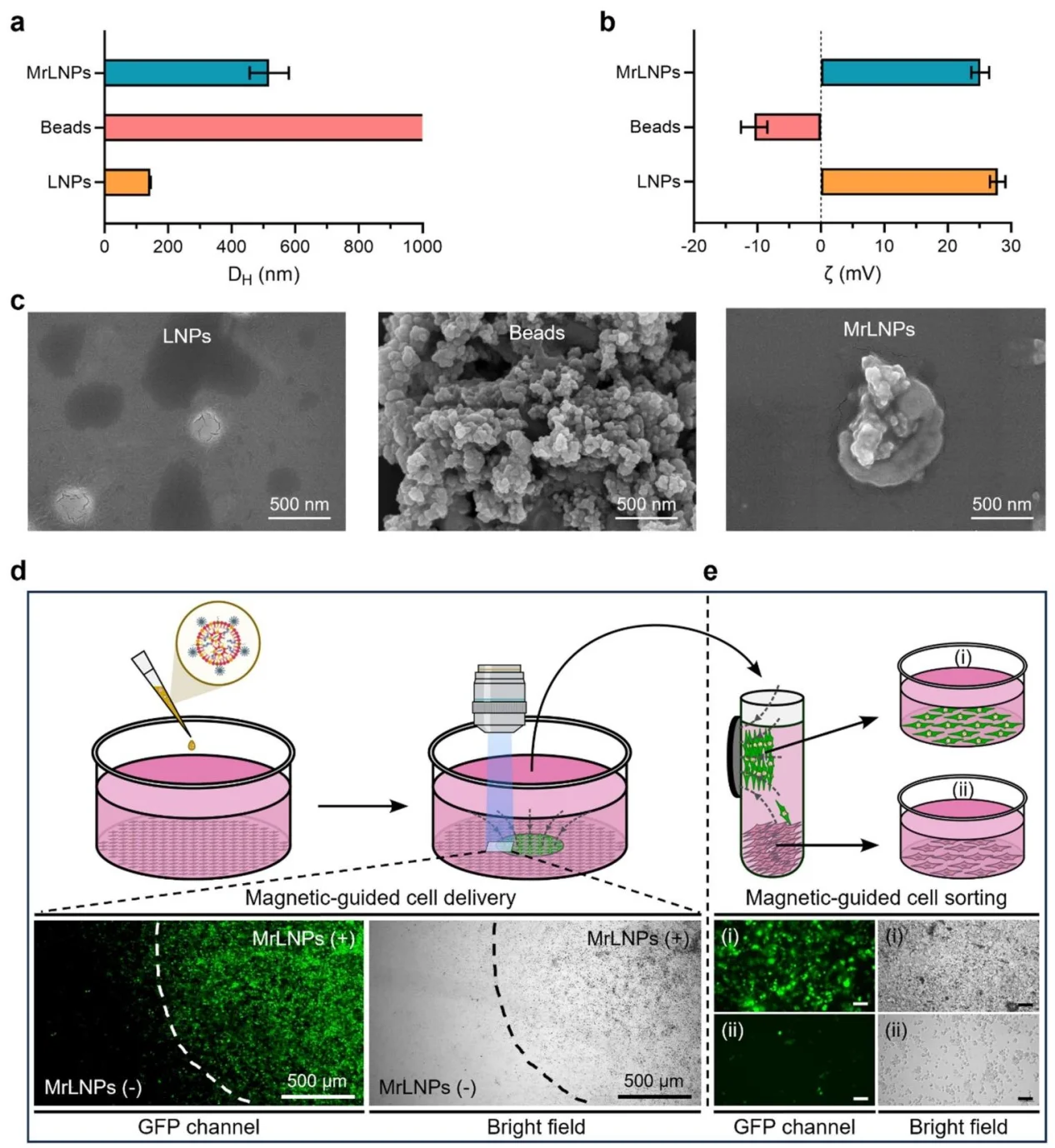
Figure 3. Physicochemical Characterization and Magnetic Functionality of MrLNPs
2. Broad-Spectrum In Vitro mRNA Delivery Capability of MrLNPs
Under external magnetic stimulation, efficient mRNA delivery was achieved within 20 minutes, with delivery performance reaching saturation during this time window. Compared with conventional LNPs, MrLNPs enhanced the delivery efficiency of FLuc mRNA, GLuc mRNA, and sFlt-1 mRNA by 6.6-fold, 2.4-fold, and 5.3-fold, respectively. Co-delivery of CRISPR-Cas9 mRNA and sgRNA also enabled highly efficient knockdown of eGFP expression, while delivery of FLuc siRNA resulted in a 26.7-fold improvement in gene silencing efficiency over conventional LNP formulations. In addition, the platform supported efficient mRNA delivery across multiple cell lines, including HeLa, THP-1, NIH/3T3, and MC38 cells, and successfully penetrated 3D cellular architectures to achieve mRNA expression throughout the entire structure. Importantly, the magnetic-responsive strategy was readily adaptable to multiple LNP systems, including TT3 LNPs and qtB-UC18 LNPs, highlighting the broad versatility of the MrLNP platform.
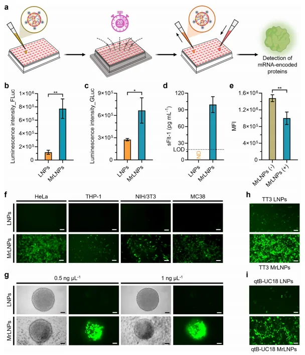
Figure 4. Intracellular mRNA Delivery Mediated by MrLNPs
3. Intracellular Trafficking and In Vitro Biocompatibility of MrLNPs
Fluorescence imaging revealed that under magnetic stimulation, MrLNPs achieved substantially higher cellular uptake within 20 minutes than conventional LNPs achieved even after 6 hours of incubation.
Mechanistic studies indicated that macropinocytosis was the dominant internalization pathway under magnetic guidance, while the contributions of clathrin-mediated and caveolae-mediated endocytosis were reduced. Colocalization analyses further confirmed that MrLNPs efficiently promoted endosomal escape, allowing mRNA payloads to be released into the cytoplasm for translation. At elevated mRNA doses, MrLNPs maintained significantly higher delivery efficiency and lower cytotoxicity compared with conventional LNPs. No obvious cytotoxicity was observed in 293T or HeLa cells at therapeutically effective mRNA doses.
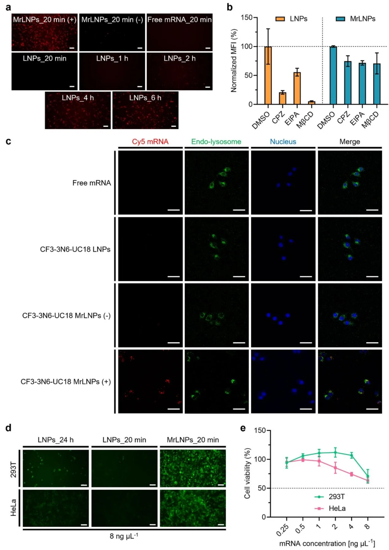
Figure 5. Cellular Uptake, Intracellular Trafficking, and Biocompatibility of MrLNPs
4. In Vivo Delivery Potency and Biosafety of MrLNPs
For the in vivo studies, N42-grade magnets were selected to provide strong magnetic stimulation. Following intraperitoneal injection of MrLNPs under an external magnetic field, bioluminescence imaging performed within 30 minutes revealed a 33.4-fold increase in signal intensity compared with the conventional LNP group. Ex vivo imaging further demonstrated markedly enhanced protein expression in multiple organs, with increases of 20.5-fold in the liver, 44.3-fold in the spleen, 5.4-fold in the lungs, and 33.8-fold in the pancreas. Even the heart and kidneys, which are hard to target, also exhibited an average 2.4-fold improvement in delivery efficiency. Importantly, in vivo biosafety evaluation showed that MrLNPs caused no detectable impairment of liver or kidney function and induced no pathological abnormalities in major organs, indicating favorable biocompatibility and biosafety.
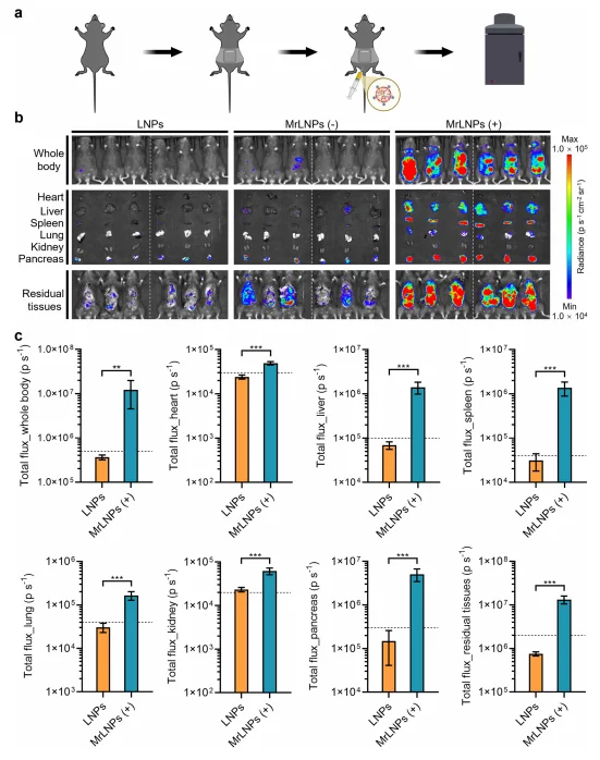
Figure 6. Rapid In Vivo mRNA Delivery Mediated by MrLNPs
Significance and Innovation
This study reports the first successful construction of magnetic responsive lipid nanoparticles (MrLNPs), overcoming the long-standing limitation of slow intracellular uptake associated with conventional mRNA delivery systems. The platform enables rapid cellular internalization within 20 minutes and achieves more than a 33-fold enhancement in in vivo delivery efficiency under magnetic stimulation. In addition to its broad delivery versatility, MrLNPs also demonstrate magnetic-guided cell sorting capability and favorable biocompatibility, supporting the delivery of multiple nucleic acid payloads, including mRNA, CRISPR-Cas9 mRNA plus sgRNA, and siRNA, across diverse cell lines and 3D culture systems. Collectively, these findings establish MrLNPs as a promising delivery platform for acute and critical diseases requiring immediate protein expression, while further expanding the translational landscape of mRNA nanomedicine.
Summary
The study presents a novel magnetic responsive lipid nanoparticle platform capable of rapid and efficient mRNA delivery both in vitro and in vivo under external magnetic stimulation. It systematically defined the optimal formulation parameters, intracellular trafficking mechanisms, and in vivo biodistribution characteristics of MrLNPs, while demonstrating their high delivery efficiency, broad applicability, and favorable safety profile. Overall, MrLNPs provide a powerful new strategy for accelerated mRNA therapeutics and hold substantial translational potential in acute disease treatment, genome engineering, vaccine delivery, and other next-generation nucleic acid therapies.
Support Provided by Ubigene
In this study, Ubigene supplied stable EGFP-expressing human embryonic kidney cells (293T-EGFP) as a quantitative and visualizable model for evaluating genome editing efficiency. These cells were used to validate the co-delivery of CRISPR-Cas9 mRNA and sgRNA mediated by MrLNPs, as well as the resulting functional gene knockout capability.
Ubigene’s stable EGFP cells feature low passage number, high viability and robust cellular condition. Constructed using a lentiviral transduction approach, these cells stably express EGFP fluorescent protein and are well suited for applications such as high-throughput drug screening, in vivo fluorescence tracing, and stable cell line development. Researchers interested in the same EGFP cell models used in this study are welcome to contact Ubigene for additional information.
Contact us to learn more>>> Promotions