Potential strategy for RA
prevention:reduced DNA fragment accumulation by upregulating TREX1
Background
Rheumatoid arthritis (RA) is a chronic and systemic autoimmune
disease with poorly understood pathogenesis. Several factors such as age, smoking history, poor body mass index
(BMI), immunogenicity and gene polymorphisms have been shown to be potentially associated with the pathogenesis
of RA and its refractoriness. As an inevitable process in life, aging is a
major contributor to the increasing risk of various diseases.
Metabolic DNA damage and sustained activation of the immune system
are important biological activities that drive cellular senescence. During cellular senescence, DNA fragments
accumulate in blood and enter the circulation to become circulating free DNA (cfDNA). Of note, DNA fragmentation
is also known to be a biological feature of many diseases, such as trauma, sepsis, aseptic inflammation,
myocardial infarction, stroke, transplant rejection, diabetes mellitus, sickle cell disease, and others. Both
Aicardi-Goutières syndrome and familial frostbite lupus share a common phenotypic feature - abnormal activation
of the immune system caused by the inability to clear self DNA or self RNA fragments in a timely manner. After
treatment with disease-modifying antirheumatic drugs (DMARDs), DNA fragmentation was significantly reduced in
the plasma of RA patients, which illustrated that DNA fragmentation may be associated with RA pathogenesis.
Based on the hypothesis that the increased incidence of RA in the middle-aged and elderly patients may be
related to age-related accumulation of DNA fragments, clarifying the source
of DNA fragments in the plasma of these patients and investigating how to improve the efficiency of the
clearance mechanism may open new avenues for the prevention and treatment strategies for RA.
To validate that DNA fragmentation is one of the factors
contributing to the development of rheumatoid arthritis, Weidan Luo's research team from Biophysics Laboratory
for Innovative Drug Discovery, State Key Laboratory of Quality Research
in Chinese Medicine, Macau University of Science and Technology, Macao, China, did a series of studies and
published an article entitled “Self-DNA
accumulation as a risk factor for accelerating the pathogenesis of rheumatoid arthritis in elderly
individuals” in Research Square. The article calls that the DNA fragments are the effective makers
of inflammation-releasing mediators, whereas the TREX1 gene is a potent degrader of DNA fragments and suggests
that TREX1 is a key gene regulating cellular immunity and aging. This conclusion provides us with a new idea to prevent rheumatoid arthritis by rapidly clearing excess DNA fragments from the
body to ensure that senescent cells remain healthy.
3'->5' DNA exonuclease
TREX1 significantly
downregulated in RA
patients
To investigate whether accumulation of DNA fragments could
stimulate an aberrant immune response, the researchers first determined the gene expression levels of TREX1 (a
DNA fragment clearance enzyme) and cGAS (a DNA fragment sensor) in peripheral blood samples from healthy
volunteers, rheumatoid arthritis (RA) patients, and osteoarthritis (OA) patients. The results in Fig.1A and Fig.1B show that the gene expression level of TREX1 but not cGAS was significantly
decreased in RA patients compared with healthy controls and OA patients, suggesting that DNA fragment accumulation is closely involved in human RA
pathogenesis. Fig. 1C demonstrates that transcript levels of both TREX1 and cGAS were
high in blood samples from healthy aged rats (>12 months old) compared with young control rats (4 weeks old).
Moreover, overexpression of TREX1, which can clear excessive DNA fragments in aged rats, can
prevent aberrant immune responses and maintain the health status of animals.
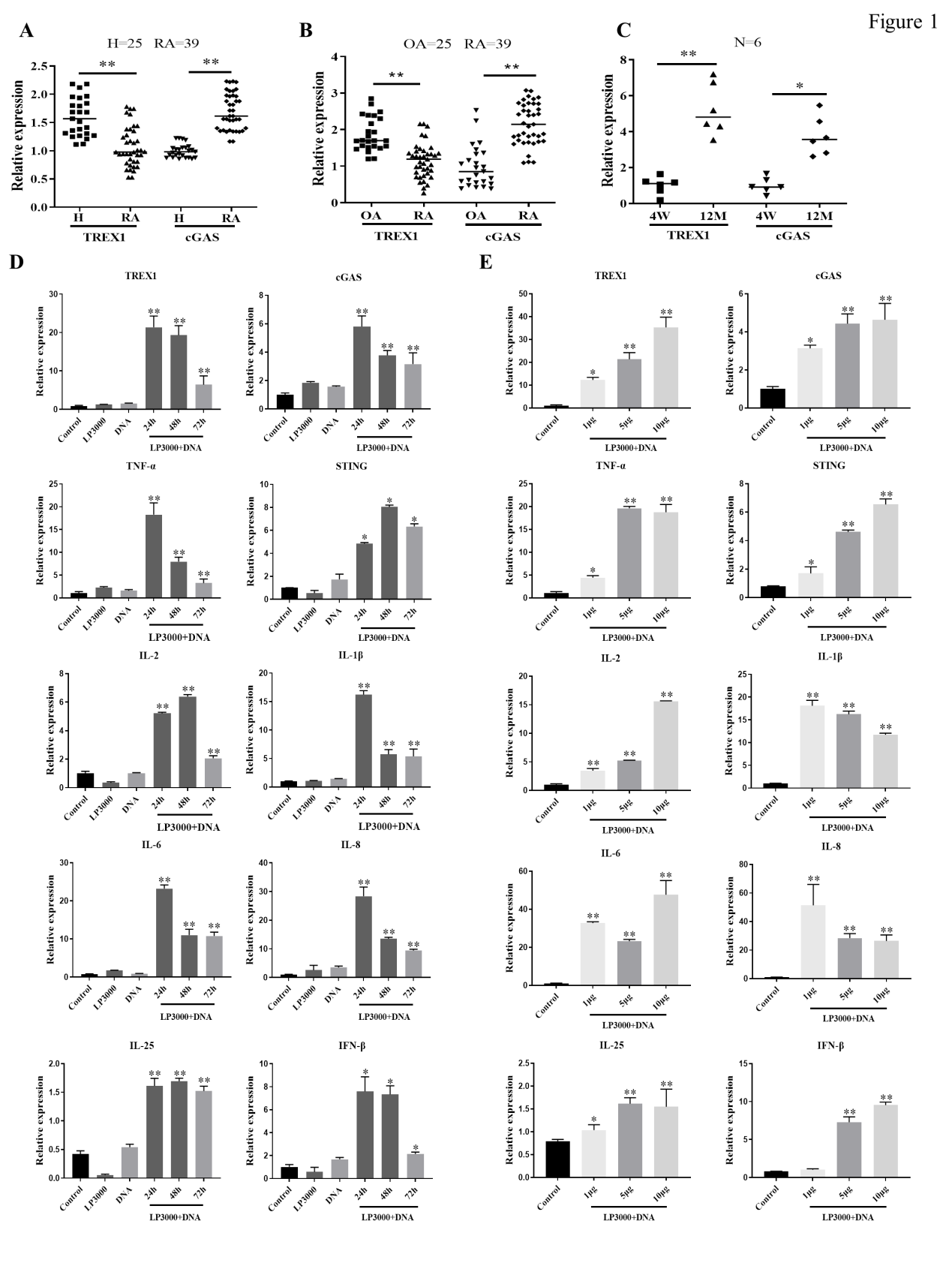
Figure 1
Role of TREX1 and cGAS signaling pathways
in DNA
fragment-induced release of proinflammatory cytokines
Transfection of DNA fragments into RA-FLSs (rheumatoid arthritis
fibroblast-like synoviocytes) stimulated the expression of the DNA fragment clearance enzyme TREX1 and
cGAS-STING, and upregulated the expression of proinflammatory cytokines (TNF-α, IL-1β, IL-2, IL-6, IL-8, IL-25
and IFN-β), and all were dose-dependent (Fig. 1D&E). However, the increased expression of TREX1, cGAS-STING,
and proinflammatory cytokines occurred only within the first 24 hours of transfection with DNA fragments, and
the expression of these activated genes declined after 48 to 72 hours of transfection. Thus, the researchers
speculate that DNA fragment-induced TREX1 upregulation may contribute to the clearance of DNA fragments that
accumulate in the cytoplasm, reducing cGAS-STING and proinflammatory cytokine levels.
Moreover, Western blot results also confirmed that TREX1 and cGAS
protein expression was time - and dose-dependent with the introduction of DNA fragments into RA-FLSs (Fig. 2A).
To reveal the roles of TREX1 and cGAS-STING signaling in DNA fragment-induced inflammatory responses, TREX1 and cGAS were silenced by siRNA in RA-FLSs before
transfection of DNA fragments,
and cGAS knockout RA-FLSs were constructed. As shown in Fig. 2B, silencing TREX1
expression activated cGAS-STING signaling, which further promoted
proinflammatory cytokine release in RA-FLSs which was transfected with DNA fragments, whereas knockout of cGAS
significantly downregulated cGAS-STING signaling and inhibited the release of DNA fragment-induced
proinflammatory cytokine. These results confirm that DNA fragments activate inflammatory responses and that
both TREX1 and cGAS play
regulatory roles in DNA fragment-induced inflammatory responses.
Gene knockout cells have now
been widely used in studies of disease treatment, signaling pathways, and target validation. Ubigene KO Cell Line Bank covers more than
thousands of genes, meeting the
three major research needs mentioned above. Over 3000 types of off-shelf KO cells are ready for 1-week delivery, as low as US$1980, helping you greatly shorten the experimental timeline. Click here to search the cell line of your interest, one-stop accelerates the experiment >>
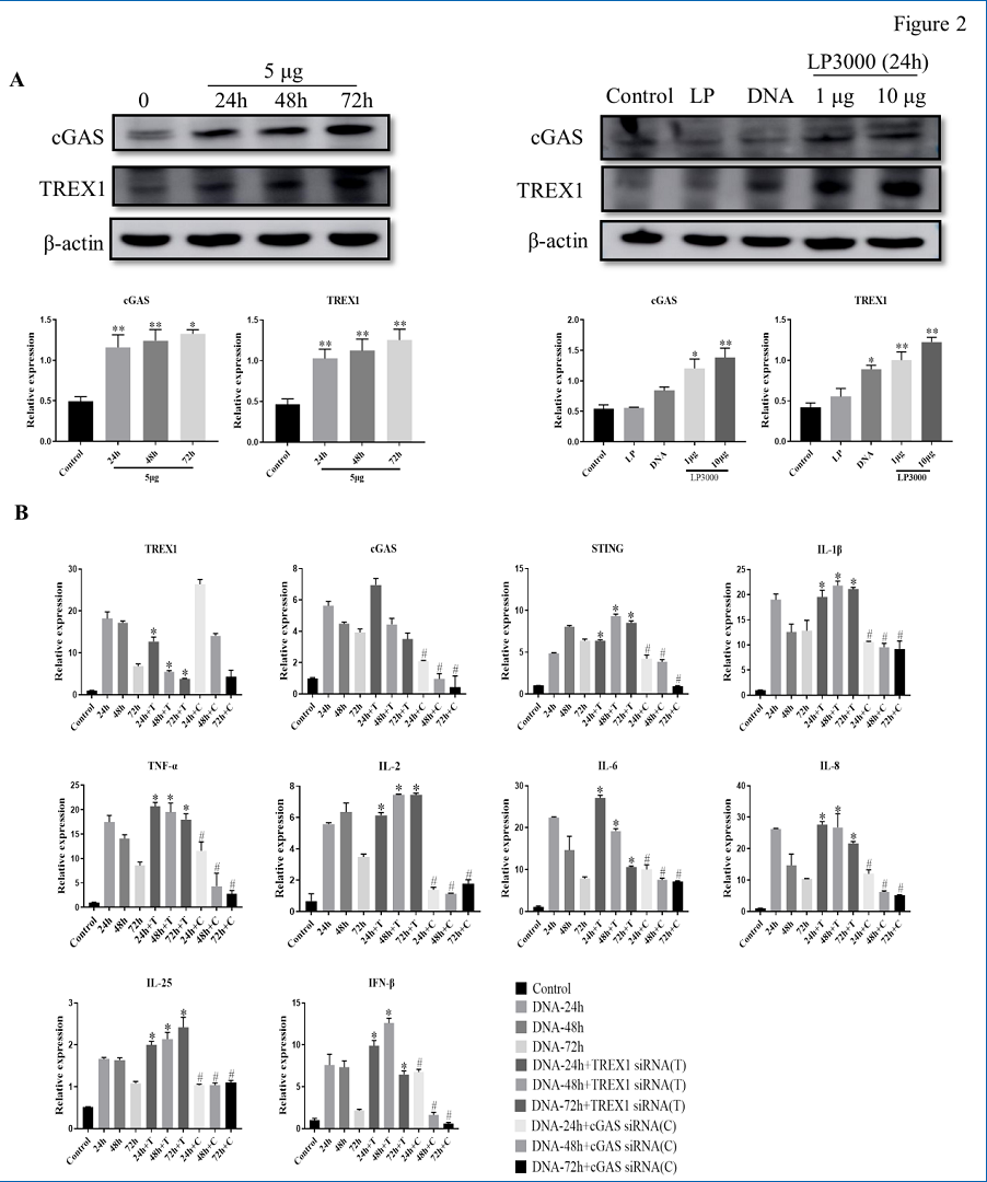
Figure 2
Joint injection of DNA fragments induced
arthritis in TREX1
conditional knockout (TREX1Cre) rats
To elucidate the role of TREX1 in the pathogenic mechanism of DNA fragmentation in
adjuvant-induced arthritis (AIA) model, the researchers established a Cre-loxP-mediated TREX1 conditional
knockout in SD rats (TREX1Cre) by CRISPR/Cas-mediated genome editing, in
which Cre adeno-associated virus was provided by
Ubigene. One of
the results in Fig. 3A-C shows after injection of 200ug DNA fragments into the hind limb joint
cavity, severe hind paw swelling, erythema, and joint rigidity were observed in
both TREX1Cre/+ AIA rats and healthy control TREX1Cre/+ rats. Moreover, TREX1 knockout or
downregulation may increase the severity of arthritis in control and AIA rats with hindlimb joint injection of
DNA fragments. These findings suggest that TREX1 may have a beneficial
therapeutic effect on RA by clearing cfDNA from joint
tissues.
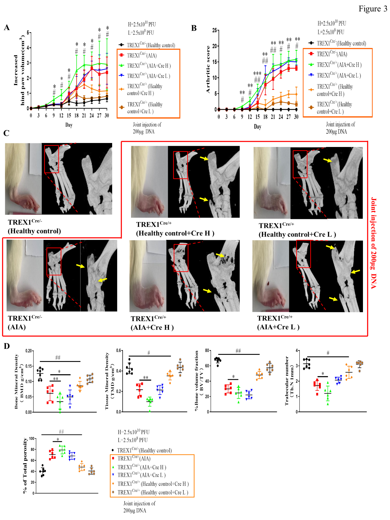
Figure 3
DNA fragments promoted T-cell activation
and reduced Treg
cell populations in TREX1Cre rats
To examine the effects of DNA fragments on autoimmune reactions in rats, sonicated rat
muscle DNA fragments were injected via the tail vein into both healthy control TREX1Cre/+ rats and AIA model TREX1Cre/+ rats. Consistent with the above experimental results, both
the healthy control TREX1Cre/+ rats and the AIA
model TREX1Cre/+ rats injected with the DNA
fragment showed more severe joint swelling in the hind limbs, significantly higher arthritis scores, and more
severe bone destruction. The results of flow cytometry analysis showed that the AIA TREX1Cre/+ model rats had significantly increased numbers of CD8 + T
lymphocytes but decreased numbers of Treg cells. After intravenous injection of DNA fragments, both AIA model TREX1Cre/+ (Cre H/L) rats and healthy control TREX1Cre/+ (Cre H/L) rats showed further increased numbers of CD8 + T
lymphocytes and decreased numbers of Treg cells compared with healthy control TREX1 wild-type rats.
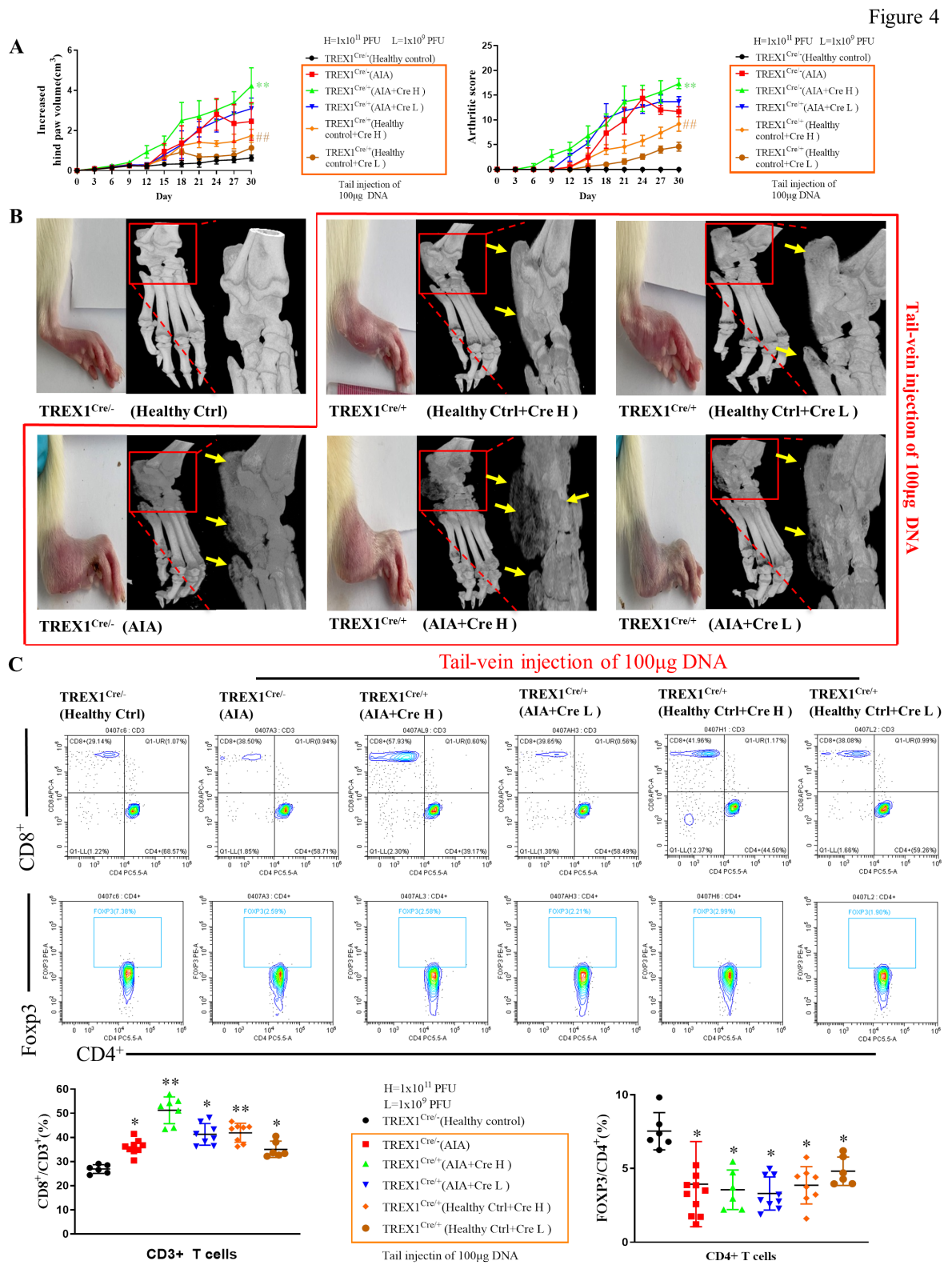
Figure 4
Adeno-associated virus (AAV)- mediated
overexpression of TREX1
suppressed synovial inflammation in AIA rats
To further investigate the role of TREX1 in the pathogenesis of AIA in rats, adeno-associated virus
expressing TREX1 (AAV-TREX1) was injected into the joint cavity
(2.5×10¹⁰ PFU or 2.5×10⁸ PFU) or tail vein (1.0×10¹¹ PFU or 1.0×10⁹ PFU) of
rats, of which the TREX1 expressing adeno-associated virus was provided by
Ubigene. The hind paws of AAV-EGFP
injected AIA rats (control) developed severe swelling, erythema, and joint rigidity. But after injection of AAV-TREX1 into the joint cavity and tail vein, AIA rats showed significant reductions in both the
arthritis score and the hind paw volume, suggesting that the severity of AIA was markedly reduced in rats with TREX1
overexpressed. So, it indicated that TREX1 may play an inhibitory role in the development of arthritis in
AIA rats by clearing DNA fragments.
Ubigene provided the
Cre and TREX1 expressing adeno-associated virus packaging service for this study, assisted in TREX1 knockout rat construction and TREX1
overexpression to verify the regulatory role of TREX1 in DNA fragment mediated inflammatory response. In
addition to adeno-associated virus, Ubigene also provides lentivirus and
adenovirus packaging services with Ubigene exclusive developed formula
which can ensure strong virus viability and high titer. And the service turnaround is fast to 3 weeks, click for more
service
details > >
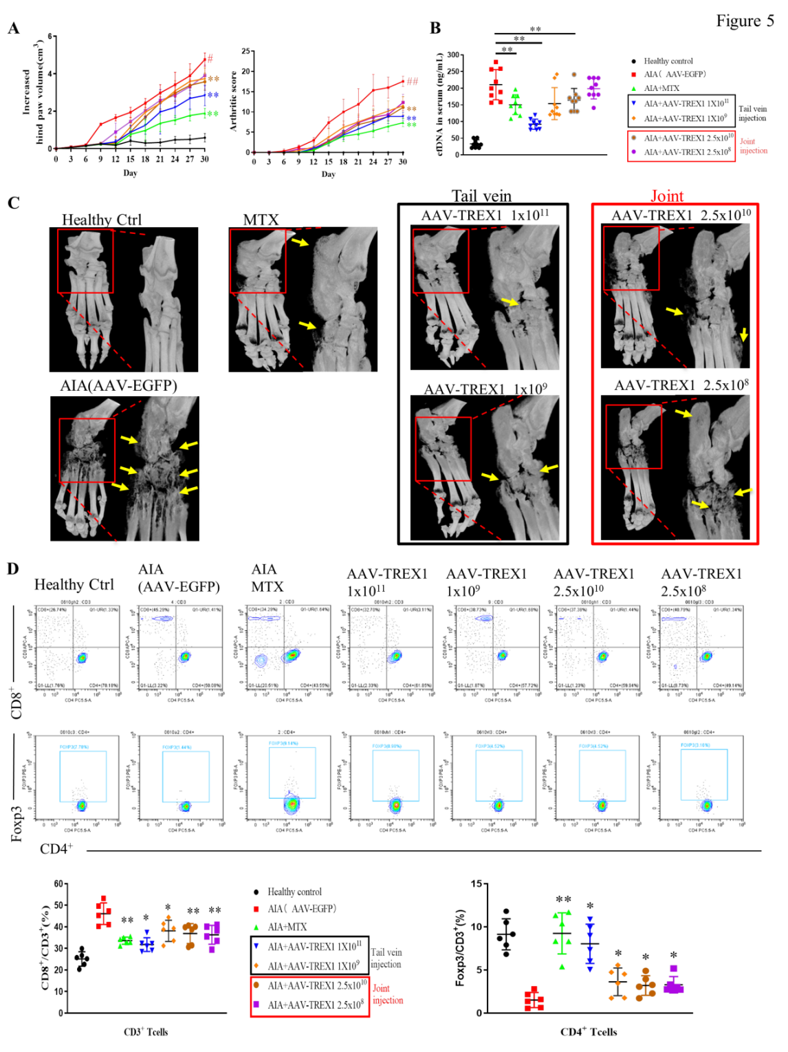
Figure 5
Aging-related cellular senescence and
arthritis led to c-Jun
/c-Fos transcription factor expression imbalance and TREX1 expression suppression
To further demonstrate the role of E2F1 in TREX1 expression, the researchers showed that E2F1 was
overexpressed in RA-FLSs in a dose-dependent manner and that the transcript levels of TREX1 and
DP-1 (important activators of E2F transcription factors) were significantly and gradually increased. In parallel,
UV-mediated DNA damage and direct DNA fragment challenge induced the expression of E2F1, DP-1, c-jun, and c-Fos,
while TREX1 was upregulated in RA-FLSs in a time-dependent manner. However, the presence of E2F1 siRNA significantly
suppressed the high TREX1 expression, indicating that E2F1 is a key transcription factor for TREX1. To further
elucidate the regulatory role of TREX1 in aging and RA, the researchers measured the expression of TREX1 and
components of the AP-1 complex (c-Jun/c-Fos) in both early- and late-passage RA-FLSs transfected with DNA fragments.
TREX1 expression was significantly lower in late-passage RA-FLSs than in early-passage RA-FLSs, regardless of
successful transfection with DNA fragments. siRNA knockdown of c-Fos significantly inhibited DNA fragment mediated
TREX1 expression in early-passage RA-FLSs. Both in aging-related cellular senescence and under arthritic
conditions, the accumulation of cfDNA can be promoted by the suppression of TREX1 transcription factors such as
E2F1 and AP-1 thereby activating the autoimmune responses.
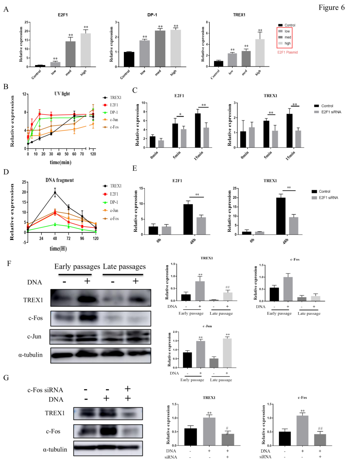
Figure 6
In summary, this study illustrates for the first
time: ①DNA fragments may be potential immunogenic factors predisposing to
RA; ②Downregulation of TREX1 will promote the release of
proinflammatory factors and may be involved in the accumulation of DNA fragments in RA patients, thereby activating the cGAS/STING signaling pathway to further
exacerbate the inflammatory response. In addition to this, the study links
autoimmune tissue inflammation to aging, delineating the efficient clearance of DNA fragments as an important
strategy for the treatment and prevention of RA and aging-related diseases.

 Subscribe Us
 Gene Editing Services
 EZ-editor™
 Red Cotton Gene knockout Project


