Latest researches of gene editing models in breast cancer

Breast cancer is one of the most common malignant tumors in women, and it has the highest incidence rate among cancers in women. CRISPR technology, as a simple, efficient and accurate gene editing method, can be used to identify drug targets and drug resistance targets, provide convenience for drug screening. It is helpful for researchers to study the drug resistance after treatment and reversal treatment. Up to now, there have plenty of publications about the research results of breast cancer using CRISPR technology. Here are some examples, hope this article can help you with your research!
Therapeutic relevance of the PP2A-B55 inhibitory kinase MASTL/Greatwall in breast cancer
PP2A is a major tumor suppressor whose inactivation is frequently found in a wide spectrum of human tumors. In particular, deletion or epigenetic silencing of genes encoding the B55 family of PP2A regulatory subunits is a common feature of breast cancer cells. A key player in the regulation of PP2A/B55 phosphatase complexes is the cell cycle kinase MASTL (also known as Greatwall). During cell division, inhibition of PP2A-B55 by MASTL is required to maintain the mitotic state, whereas inactivation of MASTL and PP2A reactivation is required for mitotic exit. MASTL as a target for human cancer therapy, in order to further understand its therapeutic relevance and its dependence on PP2A activity, teams from Cell Division and Cancer group of Spanish National Cancer Research Centre (CNIO) and Cancer Research UK Cambridge Institute used RNA interference or CRISPR/Cas9 technology to knockdown or knockout MASTL gene. The proliferation of some breast cancer cell lines, such as MDA-MB-231, was found to be impaired. The proliferative function of MASTL in these tumor cells requires its kinase activity and the presence of PP2A-B55 complexes. By using a new inducible CRISPR/Cas9 system in breast cancer cells, the genetic ablation of MASTL displays a significant therapeutic effect in vivo. This research suggests that the PP2A inhibitory kinase MASTL may have both prognostic and therapeutic value in human breast cancer.
Ubigene offers CRISPR gene knockout and RNAi gene knockdown stable cell line services. Now it is only $1780 to get a knockdown stable cell line, visit us to learn more>>

Fig. 1
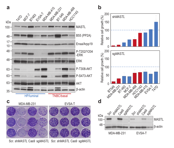
Fig. 2
Linc-RoR promotes MAPK/ERK signaling and confers estrogen-independent growth of breast cancer
The conversion from estrogen-dependent to estrogen-independent state of ER+ breast cancer cells is the key step to promote resistance to endocrine therapies. In order to study the potential mechanism of MAPK/ERK signaling pathway in the growth of estrogen independent breast cancer cells, a research team from Jiangsu University selected a group of lncRNA from the database and analyzed their expression. CRISPR/Cas9 was employed to knockout (KO) linc-RoR in MCF-7 cells, while rescue experiments were carried out to re-express linc-RoR in KO cells. Colony formation and MTT assays were used to examine the role of linc-RoR in estrogen-independent growth and tamoxifen resistance. Western blot and qRT-PCR were used to determine the change of protein and lncRNA levels, respectively. The expression of DUSP7 in clinical specimens was downloaded from Oncomine (www.oncomine.org) and the dataset from Kaplan-Meier Plotter (http://kmplot.com) was used to analyze the clinical outcomes in relation to DUSP7. They identified that linc-RoR functions as an onco-lncRNA to promote estrogen-independent growth of ER+ breast cancer. Under estrogen deprivation, linc-RoR causes the upregulation of phosphorylated MAPK/ERK pathway which in turn activates ER signaling. Knockout of linc-RoR abrogates estrogen deprivation-induced ERK activation as well as ER phosphorylation, whereas re-expression of linc-RoR restores all above phenotypes.
Moreover, they show that the ERK-specific phosphatase Dual Specificity Phosphatase 7 (DUSP7), also known as MKP-X, is involved in linc-RoR KO-induced repression of MAPK/ERK signaling. Interestingly, linc-RoR KO increases the protein stability of DUSP7, resulting in repression of ERK phosphorylation. Clinical data analysis reveal that DUSP7 expression is lower in ER+ breast cancer samples than that in ER- breast cancer. Moreover, downregulation of DUSP7 expression is associated with poor patient survival.
Taken together, these results suggest that linc-RoR promotes estrogen-independent growth and activation of MAPK/ERK pathway of breast cancer cells by regulating the ERK-specific phosphatase DUSP7. Thus, this study might help not only in establishing a role for linc-RoR in estrogen-independent and tamoxifen resistance of ER+ breast cancer, but also suggesting a link between linc-RoR and MAPK/ERK pathway.
In order to help researchers study MAPK signaling pathway, Ubigene constructed KO cells of all key genes in MAPK pathway. In addition, our KO cell bank also includes NF-kB, Hedgehog, JAK-STAT, PI3K/AKT, Notch, TGF-β, Wnt pathways. As low as $1980, deliver in 1 week. Click here to explore your KO cell lines>>
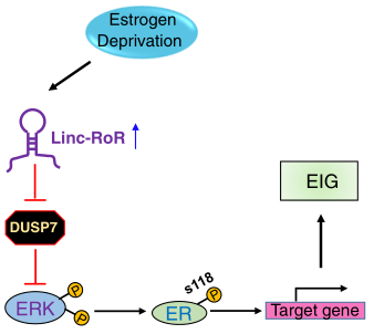
Fig. 3
Roles of acid-extruding ion transporters in regulation of breast cancer cell growth in a 3-dimensional microenvironment
Ubigene has successfully edited thousands of genes in about 200 cell lines, and successfully constructed a Knockout Cell Line Bank with nearly 2000 genes. In-stock KO cell line starting from $1980, deliver in 1 week! Click here to explore more KO cell lines>>

 Subscribe Us
 Gene Editing Services
 EZ-editor™
 Red Cotton Gene knockout Project


